• 文献检索
  • 文档翻译
  • 深度研究
  • 学术资讯
  • Suppr Zotero 插件Zotero 插件
  • 邀请有礼
  • 套餐&价格
  • 历史记录
应用&插件
Suppr Zotero 插件Zotero 插件浏览器插件Mac 客户端Windows 客户端微信小程序
定价
高级版会员购买积分包购买API积分包
服务
文献检索文档翻译深度研究API 文档MCP 服务
关于我们
关于 Suppr公司介绍联系我们用户协议隐私条款
关注我们

Suppr 超能文献

核心技术专利:CN118964589B侵权必究
粤ICP备2023148730 号-1Suppr @ 2026

文献检索

告别复杂PubMed语法,用中文像聊天一样搜索,搜遍4000万医学文献。AI智能推荐,让科研检索更轻松。

立即免费搜索

文件翻译

保留排版,准确专业,支持PDF/Word/PPT等文件格式,支持 12+语言互译。

免费翻译文档

深度研究

AI帮你快速写综述,25分钟生成高质量综述,智能提取关键信息,辅助科研写作。

立即免费体验

直肠癌新辅助治疗后患者磁共振弥散加权成像中外生性静脉侵犯和肿瘤沉积。

Extramural Venous Invasion and Tumor Deposit at Diffusion-weighted MRI in Patients after Neoadjuvant Treatment for Rectal Cancer.

机构信息

From the Departments of Radiology (T.H.K., N.G., D.D.B.B., V.P., M.J.G., N.H.), Pathology (C.F., J.S.), Surgery (H.M.T., J.G.A.), and Epidemiology and Biostatistics (J.Z., M.C.), Memorial Sloan-Kettering Cancer Center, 1275 York Ave, Box 29, New York, NY 10065.

出版信息

Radiology. 2023 Aug;308(2):e230079. doi: 10.1148/radiol.230079.

DOI:10.1148/radiol.230079
PMID:37581503
原文链接:https://pmc.ncbi.nlm.nih.gov/articles/PMC10478788/
Abstract

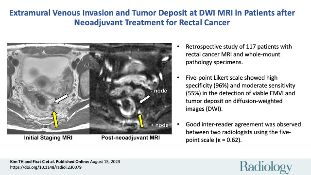
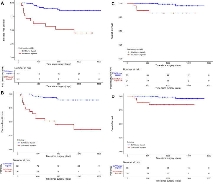
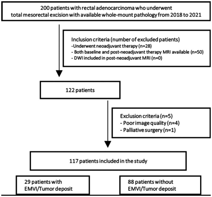
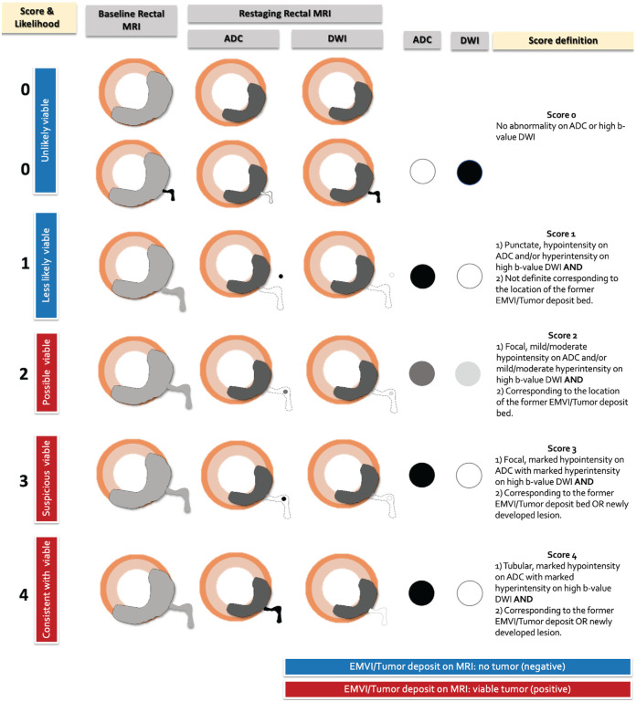
Background Diffusion-weighted (DW) imaging is useful in detecting tumor in the primary tumor bed in locally advanced rectal cancer (LARC) after neoadjuvant therapy, but its value in detecting extramural venous invasion (EMVI) and tumor deposit is not well validated. Purpose To evaluate diagnostic accuracy and association with patient prognosis of viable EMVI and tumor deposit on DW images in patients with LARC after neoadjuvant therapy using whole-mount pathology specimens. Materials and Methods This retrospective study included patients who underwent neoadjuvant therapy and surgery from 2018 to 2021. Innovative five-point Likert scale was used by two radiologists to independently evaluate the likelihood of viable EMVI and tumor deposit on restaging DW MRI scans in four axial quadrants (12 to 3 o'clock, 3 to 6 o'clock, 6 to 9 o'clock, and 9 to 12 o'clock). Diagnostic accuracy was assessed at both the per-quadrant and per-patient level, with whole-mount pathology as the reference standard. Weighted κ values for interreader agreement and Cox regression models for disease-free survival and overall survival analyses were used. Results A total of 117 patients (mean age, 56 years ± 12 [SD]; 70 male, 47 female) were included. Pathologically proven viable EMVI and tumor deposit was detected in 29 of 117 patients (25%) and in 44 of 468 quadrants (9.4%). Per-quadrant analyses showed an area under the receiver operating characteristics curve of 0.75 (95% CI: 0.68, 0.83), with sensitivity and specificity of 55% and 96%, respectively. Good interreader agreement was observed between the radiologists (κ = 0.62). Per-patient analysis showed sensitivity and specificity of 62% and 93%, respectively. The presence of EMVI and tumor deposit on restaging DW MRI scans was associated with worse disease-free survival (hazard ratio [HR], 5.6; 95% CI: 2.4, 13.3) and overall survival (HR, 8.9; 95% CI: 1.6, 48.5). Conclusion DW imaging using the five-point Likert scale showed high specificity and moderate sensitivity in the detection of viable extramural venous invasion and tumor deposits in LARC after neoadjuvant therapy, and its presence on restaging DW MRI scans is associated with worse prognosis. Published under a CC BY 4.0 license. See also the editorial by Méndez and Ayuso in this issue.

摘要

背景

扩散加权(DW)成像在局部晚期直肠癌(LARC)新辅助治疗后检测原发肿瘤床中的肿瘤方面很有用,但在检测壁外静脉侵犯(EMVI)和肿瘤沉积方面的价值尚未得到很好的验证。目的:使用全直肠标本评估 DW 图像上有活力的 EMVI 和肿瘤沉积在新辅助治疗后 LARC 患者中的诊断准确性及其与患者预后的相关性。材料与方法:本回顾性研究纳入了 2018 年至 2021 年接受新辅助治疗和手术的患者。两位放射科医生使用创新的五分 Likert 量表独立评估四个轴位象限(12 点至 3 点、3 点至 6 点、6 点至 9 点和 9 点至 12 点)再分期 DW MRI 扫描中存活的 EMVI 和肿瘤沉积的可能性。以全直肠标本为参考标准,评估象限内和患者水平的诊断准确性。使用加权κ值评估读者间的一致性,使用 Cox 回归模型评估无病生存率和总生存率。结果:共纳入 117 例患者(平均年龄 56 岁±12 岁[标准差];70 例男性,47 例女性)。29 例(25%)117 例患者和 44 例(9.4%)468 个象限中存在病理证实的有活力的 EMVI 和肿瘤沉积。象限内分析显示受试者工作特征曲线下面积为 0.75(95%CI:0.68,0.83),灵敏度和特异性分别为 55%和 96%。两位放射科医生之间观察到良好的读者间一致性(κ=0.62)。患者水平分析显示灵敏度和特异性分别为 62%和 93%。再分期 DW MRI 扫描上存在 EMVI 和肿瘤沉积与无病生存率(危险比[HR],5.6;95%CI:2.4,13.3)和总生存率(HR,8.9;95%CI:1.6,48.5)较差相关。结论:使用五分 Likert 量表的 DW 成像在检测新辅助治疗后 LARC 中的有活力的壁外静脉侵犯和肿瘤沉积方面具有高特异性和中等灵敏度,其在再分期 DW MRI 扫描上的存在与预后较差相关。在 CC BY 4.0 许可下发布。 另请参阅本期 Méndez 和 Ayuso 的社论。

https://cdn.ncbi.nlm.nih.gov/pmc/blobs/d9ee/10478788/2a8e14a36cb8/radiol.230079.fig6.jpg
https://cdn.ncbi.nlm.nih.gov/pmc/blobs/d9ee/10478788/c66428fde2e0/radiol.230079.VA.jpg
https://cdn.ncbi.nlm.nih.gov/pmc/blobs/d9ee/10478788/cb312ae233e9/radiol.230079.fig1.jpg
https://cdn.ncbi.nlm.nih.gov/pmc/blobs/d9ee/10478788/38f06a18a164/radiol.230079.fig2.jpg
https://cdn.ncbi.nlm.nih.gov/pmc/blobs/d9ee/10478788/b34b38473474/radiol.230079.fig3.jpg
https://cdn.ncbi.nlm.nih.gov/pmc/blobs/d9ee/10478788/de5e89bc2e68/radiol.230079.fig4.jpg
https://cdn.ncbi.nlm.nih.gov/pmc/blobs/d9ee/10478788/8746bb61b276/radiol.230079.fig5.jpg
https://cdn.ncbi.nlm.nih.gov/pmc/blobs/d9ee/10478788/2a8e14a36cb8/radiol.230079.fig6.jpg

相似文献

1
Extramural Venous Invasion and Tumor Deposit at Diffusion-weighted MRI in Patients after Neoadjuvant Treatment for Rectal Cancer.直肠癌新辅助治疗后患者磁共振弥散加权成像中外生性静脉侵犯和肿瘤沉积。
Radiology. 2023 Aug;308(2):e230079. doi: 10.1148/radiol.230079.
2
Diagnostic accuracy of state-of-the-art rectal MRI sequences for the diagnosis of extramural vascular invasion in locally advanced rectal cancer after preoperative chemoradiotherapy: dos or maybes?术前放化疗后局部进展期直肠癌直肠 MRI 序列对壁外血管侵犯诊断的准确性:是必然还是可能?
Eur Radiol. 2023 Oct;33(10):6852-6860. doi: 10.1007/s00330-023-09655-4. Epub 2023 Apr 28.
3
MRI of Extramural Venous Invasion in Locally Advanced Rectal Cancer: Relationship to Tumor Recurrence and Overall Survival.MRI 对局部进展期直肠癌外膜静脉侵犯的评估:与肿瘤复发和总生存的关系。
Radiology. 2018 Dec;289(3):677-685. doi: 10.1148/radiol.2018172889. Epub 2018 Aug 28.
4
Prognostic significance of MR identified EMVI, tumour deposits, mesorectal nodes and pelvic side wall disease in locally advanced rectal cancer.磁共振成像识别的包膜外血管浸润、肿瘤结节、直肠系膜淋巴结及盆腔侧壁病变在局部晚期直肠癌中的预后意义
Colorectal Dis. 2022 Apr;24(4):428-438. doi: 10.1111/codi.16032. Epub 2022 Jan 7.
5
Evaluation of Extramural Venous Invasion by Diffusion-Weighted Magnetic Resonance Imaging and Computed Tomography in Rectal Adenocarcinoma.直肠癌扩散加权磁共振成像和计算机断层扫描对外周静脉侵犯的评估。
Can Assoc Radiol J. 2019 Nov;70(4):457-465. doi: 10.1016/j.carj.2019.06.006. Epub 2019 Sep 30.
6
MRI Assessment of Extramural Venous Invasion Before and After Total Neoadjuvant Therapy for Locally Advanced Rectal Cancer and Its Association with Disease-Free and Overall Survival.局部进展期直肠癌新辅助治疗前后 MRI 评估腔外静脉侵犯及其与无病生存和总生存的关系
Ann Surg Oncol. 2023 Jul;30(7):3957-3965. doi: 10.1245/s10434-023-13225-9. Epub 2023 Mar 24.
7
Pathologic Implications of Magnetic Resonance Imaging-detected Extramural Venous Invasion of Rectal Cancer.磁共振成像检测到的直肠癌壁外静脉侵犯的病理意义
Clin Colorectal Cancer. 2023 Mar;22(1):129-135. doi: 10.1016/j.clcc.2022.10.005. Epub 2022 Nov 11.
8
Predictive value of MRI-detected extramural vascular invasion in stage T3 rectal cancer patients before neoadjuvant chemoradiation.新辅助放化疗前MRI检测壁外血管侵犯在T3期直肠癌患者中的预测价值
Diagn Interv Radiol. 2018 May-Jun;24(3):128-134. doi: 10.5152/dir.2018.17286.
9
Persistent extramural vascular invasion positivity on magnetic resonance imaging after neoadjuvant chemoradiotherapy predicts poor outcome in rectal cancer.新辅助放化疗后磁共振成像上持续的外膜血管侵犯阳性预示直肠癌预后不良。
Asian J Surg. 2021 Jun;44(6):841-847. doi: 10.1016/j.asjsur.2021.01.011. Epub 2021 Feb 8.
10
Locally advanced rectal cancer: added value of diffusion-weighted MR imaging for predicting tumor clearance of the mesorectal fascia after neoadjuvant chemotherapy and radiation therapy.局部进展期直肠癌:弥散加权磁共振成像预测新辅助放化疗后直肠系膜筋膜肿瘤退缩程度的价值。
Radiology. 2011 Sep;260(3):771-80. doi: 10.1148/radiol.11102135.

引用本文的文献

1
Extranodal extension on MRI after neoadjuvant chemoradiotherapy improves the prognostic contribution of nodal evaluation in rectal cancer.新辅助放化疗后MRI显示的结外扩展可提高淋巴结评估对直肠癌预后的预测价值。
Eur Radiol. 2025 Jul 2. doi: 10.1007/s00330-025-11788-7.
2
Diagnostic accuracy and pitfalls of MRI for restaging locally advanced rectal cancer in patients following anti-PD1 therapy plus neoadjuvant chemoradiotherapy: a multicenter study.抗PD1治疗联合新辅助放化疗后局部晚期直肠癌患者MRI再分期的诊断准确性及陷阱:一项多中心研究
Abdom Radiol (NY). 2025 Jun 17. doi: 10.1007/s00261-025-04969-x.
3
DWI of the rectum with deep learning reconstruction: comparison of PROPELLER, reduced FOV, and conventional DWI.

本文引用的文献

1
MRI Assessment of Extramural Venous Invasion Before and After Total Neoadjuvant Therapy for Locally Advanced Rectal Cancer and Its Association with Disease-Free and Overall Survival.局部进展期直肠癌新辅助治疗前后 MRI 评估腔外静脉侵犯及其与无病生存和总生存的关系
Ann Surg Oncol. 2023 Jul;30(7):3957-3965. doi: 10.1245/s10434-023-13225-9. Epub 2023 Mar 24.
2
SAR user guide to the rectal MR synoptic report (primary staging).直肠磁共振概要报告(初始分期)的SAR用户指南。
Abdom Radiol (NY). 2023 Jan;48(1):186-199. doi: 10.1007/s00261-022-03578-2. Epub 2022 Jun 26.
3
Extramural venous invasion (EMVI) revisited: a detailed analysis of various characteristics of EMVI and their role as a predictive imaging biomarker in the neoadjuvant treatment response in rectal cancer.
采用深度学习重建技术的直肠扩散加权成像:螺旋桨技术、缩小视野技术与传统扩散加权成像的比较
Abdom Radiol (NY). 2025 Apr 17. doi: 10.1007/s00261-025-04950-8.
4
Predictive value of magnetic resonance imaging parameters combined with tumor markers for rectal cancer recurrence risk after surgery.磁共振成像参数联合肿瘤标志物对直肠癌术后复发风险的预测价值
World J Gastrointest Surg. 2025 Feb 27;17(2):101897. doi: 10.4240/wjgs.v17.i2.101897.
5
Enhancing the role of MRI in rectal cancer: advances from staging to prognosis prediction.增强磁共振成像在直肠癌中的作用:从分期到预后预测的进展
Eur Radiol. 2025 Mar 6. doi: 10.1007/s00330-025-11463-x.
6
Development and evaluation of the mrTE scoring system for MRI-detected tumor deposits and extramural venous invasion in rectal cancer.直肠癌MRI检测到的肿瘤沉积物和壁外静脉侵犯的mrTE评分系统的开发与评估
Abdom Radiol (NY). 2025 Feb 15. doi: 10.1007/s00261-025-04840-z.
7
Deformable Mapping of Rectal Cancer Whole-Mount Histology with Restaging MRI at Voxel Scale: A Feasibility Study.基于体素尺度的直肠癌全直肠系膜切除术后 MRI 再分期与全直肠系膜病理学配准的可行性研究。
Radiol Imaging Cancer. 2024 Nov;6(6):e240073. doi: 10.1148/rycan.240073.
8
MRI for Rectal Cancer: Updates and Controversies- Expert Panel Narrative Review.直肠癌的磁共振成像:最新进展与争议——专家小组叙述性综述
AJR Am J Roentgenol. 2025 Jun;224(6):e2431523. doi: 10.2214/AJR.24.31523. Epub 2024 Sep 25.
9
The Role of Predictive and Prognostic MRI-Based Biomarkers in the Era of Total Neoadjuvant Treatment in Rectal Cancer.基于MRI的预测和预后生物标志物在直肠癌全新辅助治疗时代的作用
Cancers (Basel). 2024 Sep 9;16(17):3111. doi: 10.3390/cancers16173111.
10
Micro-Computed Tomography Whole-Block Imaging Reveals Origin and Path of Rectal Cancer Tumor Deposits: A Pilot Study.微计算机断层扫描全块成像揭示直肠癌肿瘤沉积物的起源和路径:一项初步研究。
Diagnostics (Basel). 2024 Aug 6;14(16):1704. doi: 10.3390/diagnostics14161704.
壁外静脉侵犯(EMVI)再探讨:对EMVI各种特征及其作为直肠癌新辅助治疗反应预测性影像生物标志物作用的详细分析
Abdom Radiol (NY). 2022 Jun;47(6):1975-1987. doi: 10.1007/s00261-022-03495-4. Epub 2022 Mar 24.
4
Interobserver variation in the classification of tumor deposits in rectal cancer-is the use of histopathological characteristics the way to go?直肠癌肿瘤沉积分类中的观察者间变异性——使用组织病理学特征是否可行?
Virchows Arch. 2021 Dec;479(6):1111-1118. doi: 10.1007/s00428-021-03197-0. Epub 2021 Sep 4.
5
Prognostic Implications of MRI-Detected EMVI and Tumor Deposits and Their Response to Neoadjuvant Therapy in cT3 and cT4 Rectal Cancer.MRI 检测的 EMVI 和肿瘤沉积及其对 cT3 和 cT4 直肠癌新辅助治疗反应的预后意义。
Int J Radiat Oncol Biol Phys. 2021 Nov 1;111(3):816-825. doi: 10.1016/j.ijrobp.2021.06.013. Epub 2021 Jun 17.
6
How to Combine Diffusion-Weighted and T2-Weighted Imaging for MRI Assessment of Pathologic Complete Response to Neoadjuvant Chemoradiotherapy in Patients with Rectal Cancer?如何将弥散加权成像和 T2 加权成像相结合,用于 MRI 评估直肠癌患者新辅助放化疗的病理完全缓解?
Korean J Radiol. 2021 Sep;22(9):1451-1461. doi: 10.3348/kjr.2020.1403. Epub 2021 May 26.
7
The prognostic value of MRI-detected extramural vascular invasion (mrEMVI) for rectal cancer patients treated with neoadjuvant therapy: a meta-analysis.MRI 检测到的直肠癌新辅助治疗患者的外膜血管侵犯(mrEMVI)的预后价值:一项荟萃分析。
Eur Radiol. 2021 Dec;31(12):8827-8837. doi: 10.1007/s00330-021-07981-z. Epub 2021 May 15.
8
MRI-Diagnosed Tumor Deposits and EMVI Status Have Superior Prognostic Accuracy to Current Clinical TNM Staging in Rectal Cancer.MRI 诊断的肿瘤沉积和 EMVI 状态对直肠癌的当前临床 TNM 分期具有更高的预后准确性。
Ann Surg. 2022 Aug 1;276(2):334-344. doi: 10.1097/SLA.0000000000004499. Epub 2020 Sep 15.
9
Utility of texture analysis on T2-weighted MR for differentiating tumor deposits from mesorectal nodes in rectal cancer patients, in a retrospective cohort.基于回顾性队列研究,T2 加权磁共振纹理分析在直肠癌患者中鉴别肿瘤沉积与直肠系膜淋巴结的效用。
Abdom Radiol (NY). 2021 Feb;46(2):459-468. doi: 10.1007/s00261-020-02653-w. Epub 2020 Jul 22.
10
Primary and post-chemoradiotherapy MRI detection of extramural venous invasion in rectal cancer: the role of diffusion-weighted imaging.直肠癌原发灶及放化疗后 MRI 检测壁外静脉侵犯:扩散加权成像的作用。
Radiol Med. 2020 Jun;125(6):522-530. doi: 10.1007/s11547-020-01137-7. Epub 2020 Feb 4.