Yao Qing-Yang, Fu Mao-Lin, Zhao Qing, Zheng Xiao-Ming, Tang Kai, Cao Li-Ming
Department of Neurology, The First Hospital of Quanzhou Affiliated to Fujian Medical University, Quanzhou 362000, Fujian Province, China.
Department of Neurology, The 910 Hospital of the Joint Logistics Support Force of the Chinese PLA, Quanzhou 362000, Fujian Province, China.
World J Clin Cases. 2023 Jul 26;11(21):5047-5055. doi: 10.12998/wjcc.v11.i21.5047.
Mechanical thrombectomy is the most effective treatment for great cerebral artery embolization within a set time window. Typically, an arteriogram does not show the localization of the stent after release and whether a thrombus is captured or not. Thus, improving the visualization of a stent in interventional therapy will be helpful for clinicians.
To analyze stent imaging findings to enhance clinicians' understanding of a special circumstance, wherein a Solitaire AB retrievable stent was visible during the imaging of a thrombus capture that improved the success rate of stent-based mechanical thrombectomy.
This was a retrospective study with four acute ischemic stroke (AIS) patients who underwent stent-based mechanical thrombectomy.
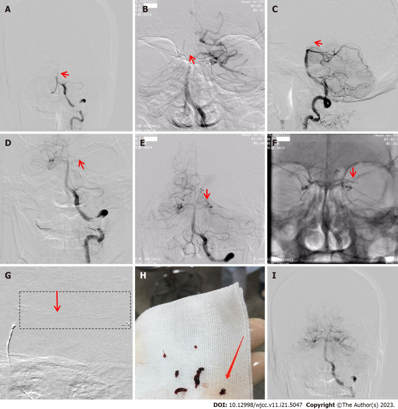
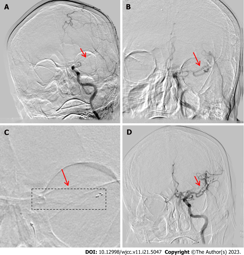
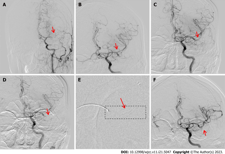
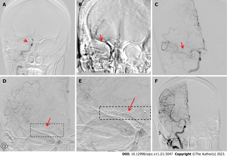
Patient 1 was a 64-year-old man admitted after 5 h of confusion; angiography revealed basilar artery occlusion. We inserted a stent into the left posterior cerebral artery-P2 segment and visualized the expanded stent that successfully captured a thrombus. Patient 2 was a 74-year-old man admitted with confusion, which lasted approximately 3 h. Angiography revealed a left middle cerebral artery (MCA)-M1 segment occlusion. A stent was deployed in the distal M2 segment, and we could visualize the stent by capturing the thrombus. Patient 3 was a 74-year-old woman admitted after experiencing left hemiplegia for 3 h. We deployed a stent at the distal right MCA-M2 segment, and the developing stent captured a large thrombus. Patient 4 was an 82-year-old man who presented with confusion for 3 h. A developing stent was placed in the distal left MCA-M1 segment, which captured a large thrombus and several fragmented thrombi.
To the best of our knowledge, this is the first report of stent imaging in patients with AIS. We demonstrated the usefulness and substantial potential of stent imaging in stent-based mechanical thrombectomy for AIS.
机械取栓术是在设定时间窗内治疗大脑大动脉栓塞最有效的方法。通常,血管造影在支架释放后无法显示其位置以及血栓是否被捕获。因此,在介入治疗中改善支架的可视化对临床医生会有帮助。
分析支架成像结果,以增强临床医生对一种特殊情况的理解,即在血栓捕获成像过程中,Solitaire AB可回收支架可见,这提高了基于支架的机械取栓术的成功率。
这是一项对4例接受基于支架的机械取栓术的急性缺血性卒中(AIS)患者的回顾性研究。
患者1为64岁男性,在意识模糊5小时后入院;血管造影显示基底动脉闭塞。我们在左大脑后动脉P2段置入一枚支架,并看到了展开的成功捕获血栓的支架。患者2为74岁男性,因意识模糊入院,持续约3小时。血管造影显示左大脑中动脉(MCA)M1段闭塞。在M2段远端置入一枚支架,通过捕获血栓我们能够看到该支架。患者3为74岁女性,在出现左侧偏瘫3小时后入院。我们在右侧MCA M2段远端置入一枚支架,展开的支架捕获了一个大血栓。患者4为82岁男性,出现意识模糊3小时。在左侧MCA M1段远端置入一枚展开的支架,该支架捕获了一个大血栓和几个破碎的血栓。
据我们所知,这是关于AIS患者支架成像的首次报告。我们证明了支架成像在AIS基于支架的机械取栓术中的有用性和巨大潜力。