• 文献检索
  • 文档翻译
  • 深度研究
  • 学术资讯
  • Suppr Zotero 插件Zotero 插件
  • 邀请有礼
  • 套餐&价格
  • 历史记录
应用&插件
Suppr Zotero 插件Zotero 插件浏览器插件Mac 客户端Windows 客户端微信小程序
定价
高级版会员购买积分包购买API积分包
服务
文献检索文档翻译深度研究API 文档MCP 服务
关于我们
关于 Suppr公司介绍联系我们用户协议隐私条款
关注我们

Suppr 超能文献

核心技术专利:CN118964589B侵权必究
粤ICP备2023148730 号-1Suppr @ 2026

文献检索

告别复杂PubMed语法,用中文像聊天一样搜索,搜遍4000万医学文献。AI智能推荐,让科研检索更轻松。

立即免费搜索

文件翻译

保留排版,准确专业,支持PDF/Word/PPT等文件格式,支持 12+语言互译。

免费翻译文档

深度研究

AI帮你快速写综述,25分钟生成高质量综述,智能提取关键信息,辅助科研写作。

立即免费体验

头皮 EEG 进行规范脑映射及其潜在的临床应用。

Normative brain mapping using scalp EEG and potential clinical application.

机构信息

CNNP Lab, Interdisciplinary Computing and Complex BioSystems Group, School of Computing, Newcastle University, Newcastle Upon Tyne, NE4 5DG, UK.

Department of Clinical and Experimental Epilepsy, UCL Queen Square Institute of Neurology, University College London, Queen Square, London, WC1N 3BG, UK.

出版信息

Sci Rep. 2023 Aug 18;13(1):13442. doi: 10.1038/s41598-023-39700-7.

DOI:10.1038/s41598-023-39700-7
PMID:37596291
原文链接:https://pmc.ncbi.nlm.nih.gov/articles/PMC10439201/
Abstract

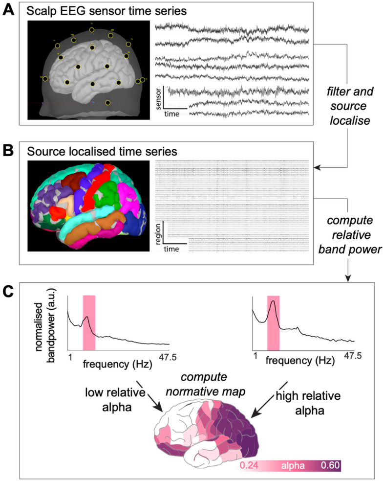
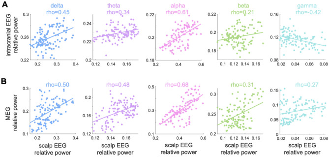
A normative electrographic activity map could be a powerful resource to understand normal brain function and identify abnormal activity. Here, we present a normative brain map using scalp EEG in terms of relative band power. In this exploratory study we investigate its temporal stability, its similarity to other imaging modalities, and explore a potential clinical application. We constructed scalp EEG normative maps of brain dynamics from 17 healthy controls using source-localised resting-state scalp recordings. We then correlated these maps with those acquired from MEG and intracranial EEG to investigate their similarity. Lastly, we use the normative maps to lateralise abnormal regions in epilepsy. Spatial patterns of band powers were broadly consistent with previous literature and stable across recordings. Scalp EEG normative maps were most similar to other modalities in the alpha band, and relatively similar across most bands. Towards a clinical application in epilepsy, we found abnormal temporal regions ipsilateral to the epileptogenic hemisphere. Scalp EEG relative band power normative maps are spatially stable across time, in keeping with MEG and intracranial EEG results. Normative mapping is feasible and may be potentially clinically useful in epilepsy. Future studies with larger sample sizes and high-density EEG are now required for validation.

摘要

正常的脑电图活动图谱可能是理解正常大脑功能和识别异常活动的有力资源。在这里,我们以相对频带功率为指标,提供了一个基于头皮 EEG 的正常脑图谱。在这项探索性研究中,我们研究了它的时间稳定性、与其他成像方式的相似性,并探索了一种潜在的临床应用。我们使用源定位静息状态头皮记录从 17 名健康对照者中构建了脑动力学的头皮 EEG 正常图谱。然后,我们将这些图谱与 MEG 和颅内 EEG 获得的图谱进行相关性分析,以研究它们的相似性。最后,我们使用正常图谱来对癫痫中的异常区域进行侧化。频带功率的空间模式与之前的文献广泛一致,并且在记录过程中是稳定的。头皮 EEG 正常图谱在 alpha 频段与其他模态最相似,并且在大多数频段上相对相似。在癫痫的临床应用中,我们发现了与致痫半球同侧的异常颞区。头皮 EEG 相对频带功率正常图谱在时间上是空间稳定的,与 MEG 和颅内 EEG 的结果一致。正常映射在癫痫中是可行的,并且可能具有潜在的临床应用价值。现在需要进行更大样本量和高密度 EEG 的进一步研究来进行验证。

https://cdn.ncbi.nlm.nih.gov/pmc/blobs/4934/10439201/b626b2a7f608/41598_2023_39700_Fig4_HTML.jpg
https://cdn.ncbi.nlm.nih.gov/pmc/blobs/4934/10439201/5802a9f5048a/41598_2023_39700_Fig1_HTML.jpg
https://cdn.ncbi.nlm.nih.gov/pmc/blobs/4934/10439201/068227e92590/41598_2023_39700_Fig2_HTML.jpg
https://cdn.ncbi.nlm.nih.gov/pmc/blobs/4934/10439201/4fe6dbaa3714/41598_2023_39700_Fig3_HTML.jpg
https://cdn.ncbi.nlm.nih.gov/pmc/blobs/4934/10439201/b626b2a7f608/41598_2023_39700_Fig4_HTML.jpg

相似文献

1
Normative brain mapping using scalp EEG and potential clinical application.头皮 EEG 进行规范脑映射及其潜在的临床应用。
Sci Rep. 2023 Aug 18;13(1):13442. doi: 10.1038/s41598-023-39700-7.
2
Normative brain mapping using scalp EEG and potential clinical application.使用头皮脑电图的规范性脑图谱及潜在临床应用。
ArXiv. 2023 Apr 6:arXiv:2304.03204v1.
3
Normative brain mapping of interictal intracranial EEG to localize epileptogenic tissue.对间期颅内 EEG 进行规范化脑图以定位致痫性组织。
Brain. 2022 Apr 29;145(3):939-949. doi: 10.1093/brain/awab380.
4
Electrocorticographic Activation Patterns of Electroencephalographic Microstates.脑电微状态的脑电皮质电图激活模式。
Brain Topogr. 2024 Mar;37(2):287-295. doi: 10.1007/s10548-023-00952-1. Epub 2023 Mar 20.
5
Identifying epileptogenic abnormality by decomposing intracranial EEG and MEG power spectra.通过分解颅内 EEG 和 MEG 功率谱来识别致痫异常。
J Neurosci Methods. 2024 Aug;408:110180. doi: 10.1016/j.jneumeth.2024.110180. Epub 2024 May 23.
6
Normative intracranial EEG maps epileptogenic tissues in focal epilepsy.规范化颅内脑电图可定位局灶性癫痫的致痫性组织。
Brain. 2022 Jun 30;145(6):1949-1961. doi: 10.1093/brain/awab480.
7
The human K-complex: Insights from combined scalp-intracranial EEG recordings.人类 K 复合波:头皮-颅内 EEG 联合记录的新见解。
Neuroimage. 2020 Jun;213:116748. doi: 10.1016/j.neuroimage.2020.116748. Epub 2020 Mar 17.
8
Alpha rhythm slowing in temporal lobe epilepsy across scalp EEG and MEG.颞叶癫痫患者头皮脑电图和脑磁图中α节律减慢。
Brain Commun. 2024 Dec 5;6(6):fcae439. doi: 10.1093/braincomms/fcae439. eCollection 2024.
9
Computer enhancement of scalp-sphenoidal ictal EEG in patients with complex partial seizures.复杂部分性发作患者头皮-蝶骨电极发作期脑电图的计算机增强处理
Electroencephalogr Clin Neurophysiol. 1988 Sep;70(3):205-19. doi: 10.1016/0013-4694(88)90081-8.
10
Increased delta power as a scalp marker of epileptic activity: a simultaneous scalp and intracranial electroencephalography study.δ 功率增加作为癫痫活动的头皮标志物:头皮和颅内脑电图同步研究。
Eur J Neurol. 2022 Jan;29(1):26-35. doi: 10.1111/ene.15106. Epub 2021 Sep 27.

引用本文的文献

1
Multi-centre normative brain mapping of intracranial EEG lifespan patterns in the human brain.人类大脑颅内脑电图寿命模式的多中心规范性脑图谱
Brain Struct Funct. 2025 Aug 21;230(7):138. doi: 10.1007/s00429-025-02988-4.
2
Distinct cortical encoding of acoustic and electrical cochlear stimulation.听觉和电刺激耳蜗的不同皮质编码。
bioRxiv. 2025 Aug 1:2025.08.01.668170. doi: 10.1101/2025.08.01.668170.
3
Harnessing electroencephalography connectomes for cognitive and clinical neuroscience.利用脑电图连接组学促进认知神经科学和临床神经科学发展

本文引用的文献

1
Interictal magnetoencephalography abnormalities to guide intracranial electrode implantation and predict surgical outcome.发作间期脑磁图异常用于指导颅内电极植入并预测手术结果。
Brain Commun. 2023 Oct 25;5(6):fcad292. doi: 10.1093/braincomms/fcad292. eCollection 2023.
2
Complementary structural and functional abnormalities to localise epileptogenic tissue.定位致痫灶的互补结构性和功能性异常。
EBioMedicine. 2023 Nov;97:104848. doi: 10.1016/j.ebiom.2023.104848. Epub 2023 Oct 27.
3
Validating MEG source imaging of resting state oscillatory patterns with an intracranial EEG atlas.
Nat Biomed Eng. 2025 Jul 23. doi: 10.1038/s41551-025-01442-4.
4
[Research on the relationship between resting-state spontaneous electroencephalography and task-evoked electroencephalography].静息态自发脑电图与任务诱发脑电图之间关系的研究
Sheng Wu Yi Xue Gong Cheng Xue Za Zhi. 2025 Jun 25;42(3):620-627. doi: 10.7507/1001-5515.202501021.
5
From Bedside to Desktop: A Data Protocol for Normative Intracranial EEG and Abnormality Mapping.从床边到桌面:一种用于规范性颅内脑电图和异常图谱的数据协议。
Bio Protoc. 2025 May 20;15(10):e5321. doi: 10.21769/BioProtoc.5321.
6
Pain and the Brain: A Systematic Review of Methods, EEG Biomarkers, Limitations, and Future Directions.疼痛与大脑:方法、脑电图生物标志物、局限性及未来方向的系统评价
Neurol Int. 2025 Mar 21;17(4):46. doi: 10.3390/neurolint17040046.
7
Alpha rhythm slowing in temporal lobe epilepsy across scalp EEG and MEG.颞叶癫痫患者头皮脑电图和脑磁图中α节律减慢。
Brain Commun. 2024 Dec 5;6(6):fcae439. doi: 10.1093/braincomms/fcae439. eCollection 2024.
8
Body Surface Potential Mapping: A Perspective on High-Density Cutaneous Electrophysiology.体表电位标测:高密度皮肤电生理学的视角
Adv Sci (Weinh). 2025 Jan;12(4):e2411087. doi: 10.1002/advs.202411087. Epub 2024 Dec 16.
9
Impact of moderate aerobic exercise on small-world topology and characteristics of brain networks after sport-related concussion: an exploratory study.运动相关性脑震荡后中等有氧运动对小世界拓扑结构和脑网络特征的影响:一项探索性研究。
Sci Rep. 2024 Oct 25;14(1):25296. doi: 10.1038/s41598-024-74474-6.
10
ANPHY-Sleep: an Open Sleep Database from Healthy Adults Using High-Density Scalp Electroencephalogram.ANPHY-Sleep:使用高密度头皮脑电图的健康成年人开放式睡眠数据库。
Sci Data. 2024 Aug 17;11(1):896. doi: 10.1038/s41597-024-03722-1.
利用颅内脑电图图谱验证静息态振荡模式的脑磁图源成像。
Neuroimage. 2023 Jul 1;274:120158. doi: 10.1016/j.neuroimage.2023.120158. Epub 2023 May 5.
4
Time in Brain: How Biological Rhythms Impact on EEG Signals and on EEG-Derived Brain Networks.大脑中的时间:生物节律如何影响脑电图信号及基于脑电图的脑网络
Front Netw Physiol. 2021 Sep 27;1:755016. doi: 10.3389/fnetp.2021.755016. eCollection 2021.
5
MEG abnormalities and mechanisms of surgical failure in neocortical epilepsy.MEG 异常与新皮层癫痫手术失败的机制。
Epilepsia. 2023 Mar;64(3):692-704. doi: 10.1111/epi.17503. Epub 2023 Jan 24.
6
The Theta Paradox: 4-8 Hz EEG Oscillations Reflect Both Sleep Pressure and Cognitive Control.Theta 悖论:4-8 Hz 的脑电振荡既反映睡眠压力又反映认知控制。
J Neurosci. 2022 Nov 9;42(45):8569-8586. doi: 10.1523/JNEUROSCI.1063-22.2022. Epub 2022 Oct 6.
7
The natural frequencies of the resting human brain: An MEG-based atlas.静息态人脑的固有频率:基于 MEG 的图谱。
Neuroimage. 2022 Sep;258:119373. doi: 10.1016/j.neuroimage.2022.119373. Epub 2022 Jun 11.
8
Normative intracranial EEG maps epileptogenic tissues in focal epilepsy.规范化颅内脑电图可定位局灶性癫痫的致痫性组织。
Brain. 2022 Jun 30;145(6):1949-1961. doi: 10.1093/brain/awab480.
9
Normative brain mapping of interictal intracranial EEG to localize epileptogenic tissue.对间期颅内 EEG 进行规范化脑图以定位致痫性组织。
Brain. 2022 Apr 29;145(3):939-949. doi: 10.1093/brain/awab380.
10
Quantitative Electroencephalogram Standardization: A Sex- and Age-Differentiated Normative Database.定量脑电图标准化:一个按性别和年龄区分的标准数据库。
Front Neurosci. 2021 Dec 17;15:766781. doi: 10.3389/fnins.2021.766781. eCollection 2021.