Yang Bin, Gao Yeqi, Lu Jie, Wang Yefu, Wu Ren, Shen Jie, Ren Jialiang, Wu Feiyun, Xu Hai
Department of Radiology, The First Affiliated Hospital of Nanjing Medical University, Nanjing, China.
Department of Medical Imaging, Jinling Hospital, Nanjing Medical University, Nanjing, China.
Front Oncol. 2023 Aug 4;13:1212608. doi: 10.3389/fonc.2023.1212608. eCollection 2023.
In this study, we developed and validated machine learning (ML) models by combining radiomic features extracted from magnetic resonance imaging (MRI) with clinicopathological factors to assess pulmonary nodule classification for benign malignant diagnosis.
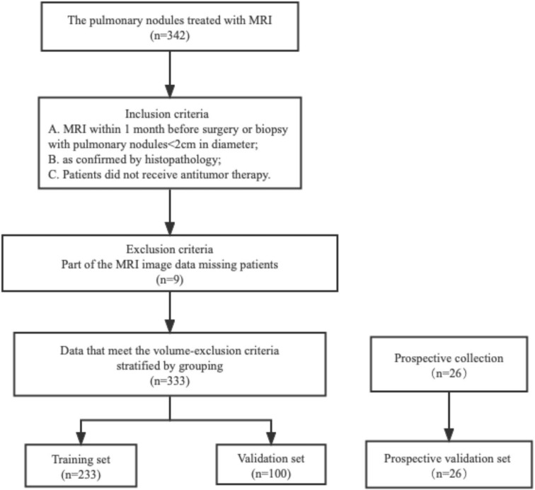
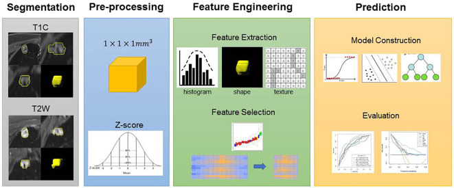
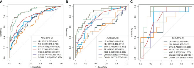
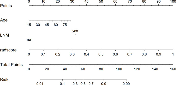
A total of 333 consecutive patients with pulmonary nodules (233 in the training cohort and 100 in the validation cohort) were enrolled. A total of 2,824 radiomic features were extracted from the MRI images (CE T1w and T2w). Logistic regression (LR), Naïve Bayes (NB), support vector machine (SVM), random forest (RF), and extreme gradient boosting (XGBoost) classifiers were used to build the predictive models, and a radiomics score (Rad-score) was obtained for each patient after applying the best prediction model. Clinical factors and Rad-scores were used jointly to build a nomogram model based on multivariate logistic regression analysis, and the diagnostic performance of the five prediction models was evaluated using the area under the receiver operating characteristic curve (AUC).
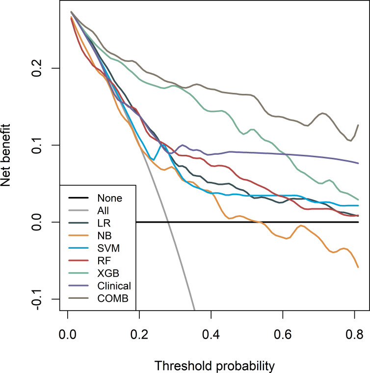
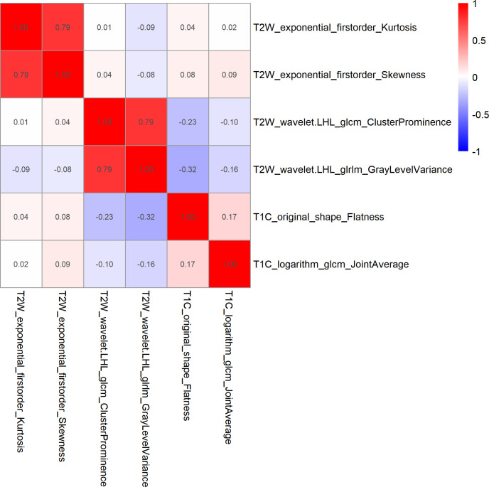
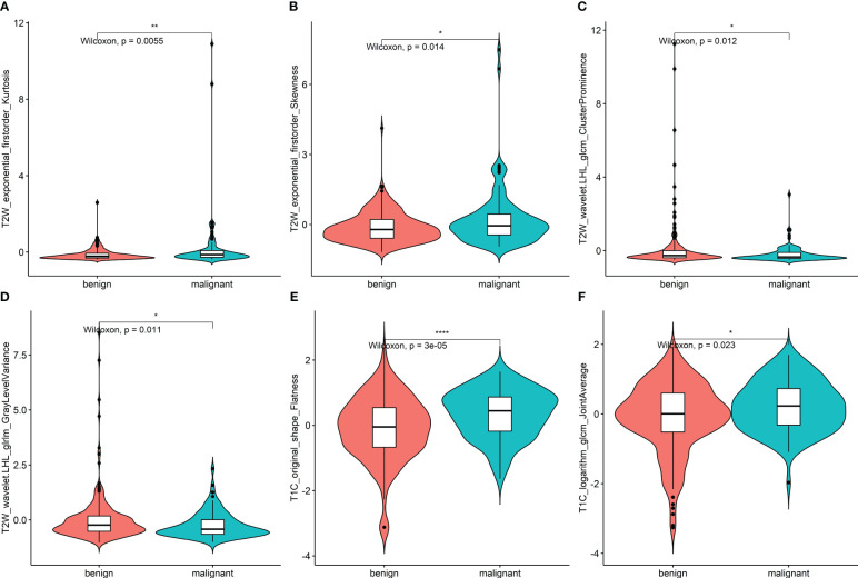
A total of 161 women (48.35%) and 172 men (51.65%) with pulmonary nodules were enrolled. Six important features were selected from the 2,145 radiomic features extracted from CE T1w and T2w images. The XGBoost classifier model achieved the highest discrimination performance with AUCs of 0.901, 0.906, and 0.851 in the training, validation, and test cohorts, respectively. The nomogram model improved the performance with AUC values of 0.918, 0.912, and 0.877 in the training, validation, and test cohorts, respectively.
MRI radiomic ML models demonstrated good nodule classification performance with XGBoost, which was superior to that of the other four models. The nomogram model achieved higher performance with the addition of clinical information.
在本研究中,我们通过将从磁共振成像(MRI)中提取的放射组学特征与临床病理因素相结合,开发并验证了机器学习(ML)模型,以评估肺结节的良恶性分类用于诊断。
共纳入333例连续的肺结节患者(训练队列233例,验证队列100例)。从MRI图像(CE T1w和T2w)中提取了总共2824个放射组学特征。使用逻辑回归(LR)、朴素贝叶斯(NB)、支持向量机(SVM)、随机森林(RF)和极端梯度提升(XGBoost)分类器构建预测模型,并在应用最佳预测模型后为每位患者获得放射组学评分(Rad-score)。基于多变量逻辑回归分析,联合临床因素和Rad-scores构建列线图模型,并使用受试者操作特征曲线(AUC)下面积评估五个预测模型的诊断性能。
共纳入161例女性(48.35%)和172例男性(51.65%)肺结节患者。从CE T1w和T2w图像中提取的2145个放射组学特征中选择了六个重要特征。XGBoost分类器模型在训练、验证和测试队列中的AUC分别为0.901、0.906和0.851,具有最高的区分性能。列线图模型在训练、验证和测试队列中的AUC值分别为0.918、0.912和0.877,提高了性能。
MRI放射组学ML模型在XGBoost下表现出良好的结节分类性能,优于其他四个模型。列线图模型在添加临床信息后性能更高。