Gao Shenghan, Chen Bo, Gao Min, Xu Yue, Yang Xueyi, Yang Chun, Pan Shaoxia
Department of Prosthodontics, Peking University School and Hospital of Stomatology & National Center for Stomatology & National Clinical Research Center for Oral Diseases & National Engineering Research Center of Oral Biomaterials and Digital Medical Devices, Beijing Key Laboratory of Digital Stomatology, Research Center of Engineering and Technology for Computerized Dentistry Ministry of Health, NMPA Key Laboratory for Dental Materials, Central Laboratory, Peking University School and Hospital of Stomatology, No. 22, Zhongguancun South Avenue, Haidian District, Beijing 100081, China.
Department of Implantology, Peking University School and Hospital of Stomatology & National Center for Stomatology & National Clinical Research Center for Oral Diseases & National Engineering Research Center of Oral Biomaterials and Digital Medical Devices, No. 22, Zhongguancun South Avenue, Haidian District, Beijing 100081, China.
Biomimetics (Basel). 2023 Aug 4;8(4):344. doi: 10.3390/biomimetics8040344.
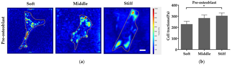
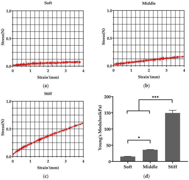
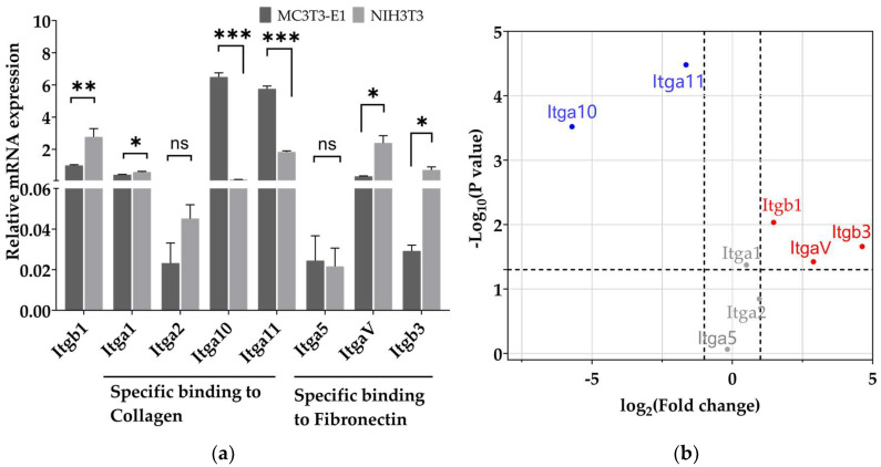
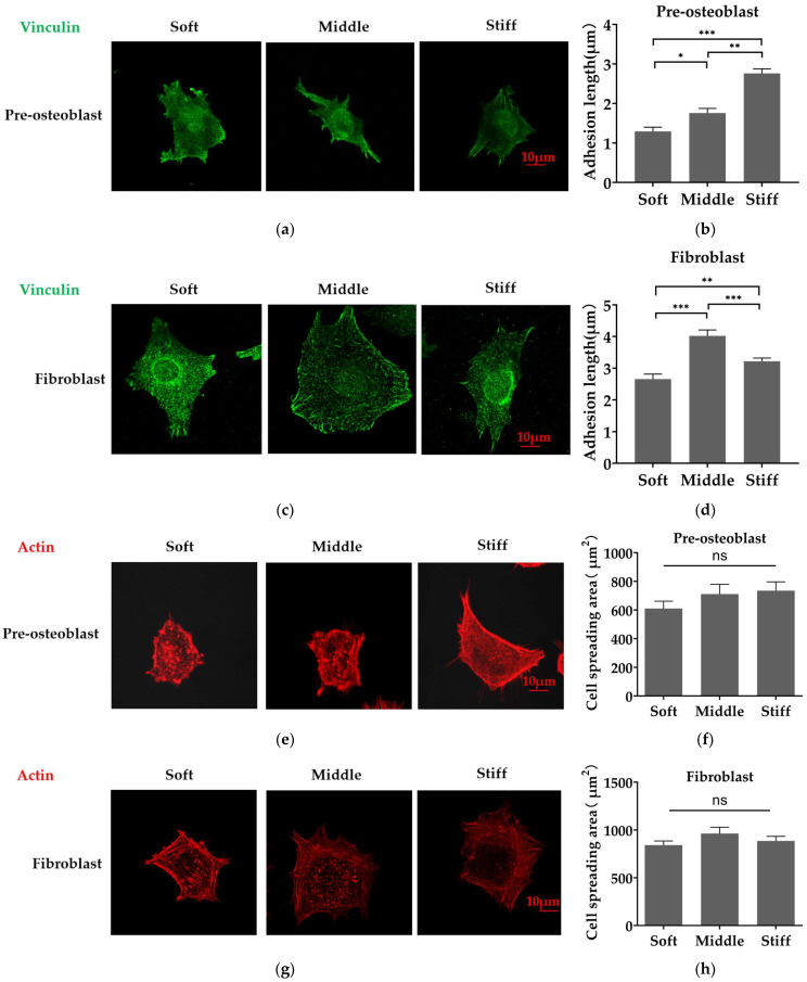
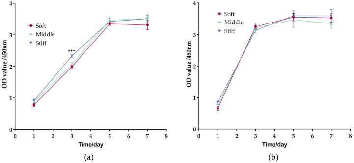
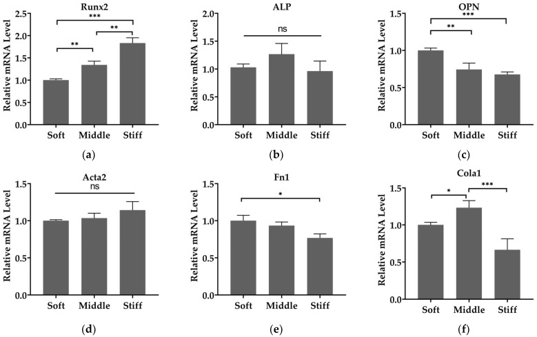
The formation of bone in a bone defect is accomplished by osteoblasts, while the over activation of fibroblasts promotes fibrosis. However, it is not clear how the extracellular matrix stiffness of the bone-regeneration microenvironment affects the function of osteoblasts and fibroblasts. This study aim to investigate the effect of bone-regeneration microenvironment stiffness on cell adhesion, cell proliferation, cell differentiation, synthesizing matrix ability and its potential mechanisms in mechanotransduction, in pre-osteoblasts and fibroblasts. Polyacrylamide substrates mimicking the matrix stiffness of different stages of the bone-healing process (15 kPa, mimic granulation tissue; 35 kPa, mimic osteoid; 150 kPa, mimic calcified bone matrix) were prepared. Mouse pre-osteoblasts MC3T3-E1 and mouse fibroblasts NIH3T3 were plated on three types of substrates, respectively. There were significant differences in the adhesion of pre-osteoblasts and fibroblasts on different polyacrylamide substrates. Runx2 expression increased with increasing substrate stiffness in pre-osteoblasts, while no statistical differences were found in the Acta2 expression in fibroblasts on three substrates. OPN expression in pre-osteoblasts, as well as Fn1 and Col1a1 expression in fibroblasts, decreased with increasing stiffness. The difference between the cell traction force generated by pre-osteoblasts and fibroblasts on substrates was also found. Our results indicated that substrate stiffness is a potent regulator of pre-osteoblasts and fibroblasts with the ability of promoting osteogenic differentiation of pre-osteoblasts, while having no effect on myofibroblast differentiation of fibroblasts.
骨缺损处的骨形成是由成骨细胞完成的,而成纤维细胞的过度活化会促进纤维化。然而,骨再生微环境的细胞外基质硬度如何影响成骨细胞和成纤维细胞的功能尚不清楚。本研究旨在探讨骨再生微环境硬度对前成骨细胞和成纤维细胞的细胞黏附、细胞增殖、细胞分化、合成基质能力及其机械转导潜在机制的影响。制备了模拟骨愈合过程不同阶段基质硬度的聚丙烯酰胺底物(15 kPa,模拟肉芽组织;35 kPa,模拟类骨质;150 kPa,模拟钙化骨基质)。将小鼠前成骨细胞MC3T3-E1和小鼠成纤维细胞NIH3T3分别接种在三种类型的底物上。前成骨细胞和成纤维细胞在不同聚丙烯酰胺底物上的黏附存在显著差异。在前成骨细胞中,Runx2表达随底物硬度增加而增加,而在三种底物上成纤维细胞的Acta2表达未发现统计学差异。前成骨细胞中的OPN表达以及成纤维细胞中的Fn1和Col1a1表达随硬度增加而降低。还发现了前成骨细胞和成纤维细胞在底物上产生的细胞牵引力差异。我们的结果表明,底物硬度是前成骨细胞和成纤维细胞的有效调节因子,具有促进前成骨细胞成骨分化的能力,而对成纤维细胞的肌成纤维细胞分化没有影响。