• 文献检索
  • 文档翻译
  • 深度研究
  • 学术资讯
  • Suppr Zotero 插件Zotero 插件
  • 邀请有礼
  • 套餐&价格
  • 历史记录
应用&插件
Suppr Zotero 插件Zotero 插件浏览器插件Mac 客户端Windows 客户端微信小程序
定价
高级版会员购买积分包购买API积分包
服务
文献检索文档翻译深度研究API 文档MCP 服务
关于我们
关于 Suppr公司介绍联系我们用户协议隐私条款
关注我们

Suppr 超能文献

核心技术专利:CN118964589B侵权必究
粤ICP备2023148730 号-1Suppr @ 2026

文献检索

告别复杂PubMed语法,用中文像聊天一样搜索,搜遍4000万医学文献。AI智能推荐,让科研检索更轻松。

立即免费搜索

文件翻译

保留排版,准确专业,支持PDF/Word/PPT等文件格式,支持 12+语言互译。

免费翻译文档

深度研究

AI帮你快速写综述,25分钟生成高质量综述,智能提取关键信息,辅助科研写作。

立即免费体验

上颌窦内安德伍德隔及其相关因素的CBCT回顾性研究——一种分类方案

The CBCT Retrospective Study on Underwood Septa and Their Related Factors in Maxillary Sinuses-A Proposal of Classification.

作者信息

Nelke Kamil, Diakowska Dorota, Morawska-Kochman Monika, Janeczek Maciej, Pasicka Edyta, Łukaszewski Marceli, Żak Krzysztof, Nienartowicz Jan, Dobrzyński Maciej

机构信息

Maxillo-Facial Surgery Ward, EMC Hospital, Pilczycka 144, 54-144 Wrocław, Poland.

Academy of Applied Sciences, Health Department, Academy of Silesius in Wałbrzych, Zamkowa 4, 58-300 Wałbrzych, Poland.

出版信息

J Pers Med. 2023 Aug 14;13(8):1258. doi: 10.3390/jpm13081258.

DOI:10.3390/jpm13081258
PMID:37623508
原文链接:https://pmc.ncbi.nlm.nih.gov/articles/PMC10455419/
Abstract

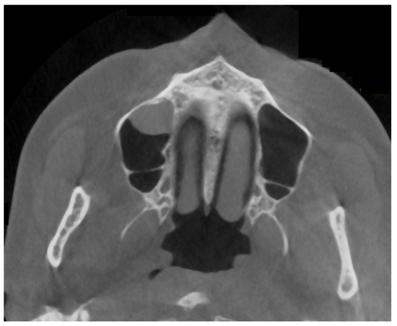
The presence of bone septum in the maxillary sinus is one of the most common anatomical findings. So-called Underwood septa (US) are an atypical bone formation in the maxillary sinuses. Mostly they are quite easily found in CBCT studies and have major importance in sinus lift procedures in dental surgery. Furthermore, the shape, location, and size of the bony septa are important in each maxillary sinus surgery. A retrospective study of 120CBCT scans from the authors' own database was conducted. Approximately 37.5% of each CBCT was associated with the occurrence of US, while just 25% had a full septum, and a total of only 14 patients had a half septa. More females have US, while healthy pneumatized maxillary sinus is most commonly found (82.22%). There is no correlation between the occurrence of silent sinus syndrome ( = 0.174), mucosal thickening ( = 0.325), or retention cyst formation ( = 0.272). Most sinuses are without any opacification in CBCT evaluation (91.11%), while other syndromes are not statistically relevant. It seems that the occurrence of Underwood septa is not statistically related to any clinical, radiological, or pathological condition within the sinus ( > 0.05). Furthermore, a more full or partial appearance of US was found in female patients.

摘要

上颌窦中骨隔的存在是最常见的解剖学发现之一。所谓的安德伍德骨隔(US)是上颌窦中的一种非典型骨形成。在锥形束计算机断层扫描(CBCT)研究中,它们大多很容易被发现,并且在牙科手术的窦底提升术中具有重要意义。此外,骨隔的形状、位置和大小在每例上颌窦手术中都很重要。对作者自己数据库中的120例CBCT扫描进行了回顾性研究。大约37.5%的CBCT与安德伍德骨隔的出现有关,而只有25%有完整的骨隔,总共只有14例患者有半骨隔。女性中安德伍德骨隔更为常见,而健康的气化良好的上颌窦最为常见(82.22%)。无症状性窦综合征的发生(=0.174)、黏膜增厚(=0.325)或潴留囊肿形成(=0.272)之间无相关性。在CBCT评估中,大多数鼻窦没有任何混浊(91.11%),而其他综合征无统计学意义。似乎安德伍德骨隔的发生与窦内的任何临床、放射学或病理状况均无统计学相关性(>0.05)。此外,在女性患者中发现安德伍德骨隔的表现更完整或更部分。

https://cdn.ncbi.nlm.nih.gov/pmc/blobs/de89/10455419/eaba5f2d6f64/jpm-13-01258-g010.jpg
https://cdn.ncbi.nlm.nih.gov/pmc/blobs/de89/10455419/e94f6a85601c/jpm-13-01258-g001.jpg
https://cdn.ncbi.nlm.nih.gov/pmc/blobs/de89/10455419/57df27200a20/jpm-13-01258-g002.jpg
https://cdn.ncbi.nlm.nih.gov/pmc/blobs/de89/10455419/d52d21c533db/jpm-13-01258-g003.jpg
https://cdn.ncbi.nlm.nih.gov/pmc/blobs/de89/10455419/090a2e63bfda/jpm-13-01258-g004.jpg
https://cdn.ncbi.nlm.nih.gov/pmc/blobs/de89/10455419/a4453419dcb0/jpm-13-01258-g005.jpg
https://cdn.ncbi.nlm.nih.gov/pmc/blobs/de89/10455419/e66a3dcc68d5/jpm-13-01258-g006.jpg
https://cdn.ncbi.nlm.nih.gov/pmc/blobs/de89/10455419/dc21d0ae6a31/jpm-13-01258-g007.jpg
https://cdn.ncbi.nlm.nih.gov/pmc/blobs/de89/10455419/8462aeb59941/jpm-13-01258-g008.jpg
https://cdn.ncbi.nlm.nih.gov/pmc/blobs/de89/10455419/0f403aa3be07/jpm-13-01258-g009.jpg
https://cdn.ncbi.nlm.nih.gov/pmc/blobs/de89/10455419/eaba5f2d6f64/jpm-13-01258-g010.jpg

相似文献

1
The CBCT Retrospective Study on Underwood Septa and Their Related Factors in Maxillary Sinuses-A Proposal of Classification.上颌窦内安德伍德隔及其相关因素的CBCT回顾性研究——一种分类方案
J Pers Med. 2023 Aug 14;13(8):1258. doi: 10.3390/jpm13081258.
2
Prevalence of bony septa, antral pathology, and dimensions of the maxillary sinus from a sinus augmentation perspective: A retrospective cone-beam computed tomography study.从鼻窦增高术角度看骨间隔、窦腔病变及上颌窦尺寸的患病率:一项回顾性锥形束计算机断层扫描研究
Imaging Sci Dent. 2016 Jun;46(2):109-15. doi: 10.5624/isd.2016.46.2.109. Epub 2016 Jun 23.
3
Evaluation of Maxillary Sinus Septa Using Cone Beam Computed Tomography (CBCT): A Retrospective Study.使用锥形束计算机断层扫描(CBCT)对上颌窦隔进行评估:一项回顾性研究。
Cureus. 2024 Aug 29;16(8):e68157. doi: 10.7759/cureus.68157. eCollection 2024 Aug.
4
Retrospective analysis of pathological changes in the maxillary sinus with CBCT.CBCT 上颌窦病变的回顾性分析。
Sci Rep. 2024 Jul 5;14(1):15529. doi: 10.1038/s41598-024-66527-7.
5
CBCT Study of Morphologic Variations of Maxillary Sinus Septa in Relevance to Sinus Augmentation Procedures.与上颌窦提升术相关的上颌窦间隔形态变异的锥形束计算机断层扫描研究
Ann Maxillofac Surg. 2020 Jan-Jun;10(1):51-56. doi: 10.4103/ams.ams_141_19. Epub 2020 Jun 8.
6
Prevalence and Characteristics of the Maxillary Sinus Septa in a Saudi Arabian Sub-population: A Retrospective Cone Beam Computed Tomography (CBCT)-Based Study.沙特阿拉伯亚人群中上颌窦隔的患病率及特征:一项基于锥形束计算机断层扫描(CBCT)的回顾性研究。
Cureus. 2023 Oct 24;15(10):e47605. doi: 10.7759/cureus.47605. eCollection 2023 Oct.
7
Mapping Maxillary Sinus Septa in a Lebanese Sample: a Radio-anatomical Study.黎巴嫩样本中上颌窦隔的影像学解剖研究
Acta Inform Med. 2020 Sep;28(3):214-218. doi: 10.5455/aim.2020.28.214-218.
8
Evaluating Maxillary Sinus Septa Using Cone Beam Computed Tomography: Is There a Difference in Frequency and Type Between the Dentate and Edentulous Posterior Maxilla?应用锥形束 CT 评估上颌窦间隔:有牙和无牙后上颌窦的频率和类型是否存在差异?
Int J Oral Maxillofac Implants. 2017 Nov/Dec;32(6):1324-1332. doi: 10.11607/jomi.5854.
9
Prevalence and morphological variations of maxillary sinus septa in different age groups: a CBCT analysis.不同年龄组上颌窦间隔的流行率和形态学变化:CBCT 分析。
Ann Saudi Med. 2020 May-Jun;40(3):200-206. doi: 10.5144/0256-4947.2020.200. Epub 2020 Jun 4.
10
Prevalence and anatomical variations of maxillary sinus septa: A cone-beam computed tomography analysis.上颌窦隔的患病率及解剖变异:锥形束计算机断层扫描分析
J Clin Exp Dent. 2022 Sep 1;14(9):e689-e693. doi: 10.4317/jced.59599. eCollection 2022 Sep.

引用本文的文献

1
What are the radiographic prevalence and anatomical patterns of maxillary sinus septa in a Turkish cohort?土耳其人群中上颌窦隔的影像学患病率及解剖模式是什么?
BMC Oral Health. 2025 May 31;25(1):862. doi: 10.1186/s12903-025-06268-9.
2
Association between sinus septa and lateral wall thickness with risk of perforation during maxillary sinus lift surgery: A systematic review and meta-analysis.上颌窦提升术中窦间隔及侧壁厚度与穿孔风险的相关性:一项系统评价与Meta分析
PLoS One. 2024 Aug 8;19(8):e0308166. doi: 10.1371/journal.pone.0308166. eCollection 2024.

本文引用的文献

1
The Use of CBCT in Evaluating the Health and Pathology of the Maxillary Sinus.锥形束计算机断层扫描(CBCT)在上颌窦健康与病变评估中的应用
Diagnostics (Basel). 2022 Nov 16;12(11):2819. doi: 10.3390/diagnostics12112819.
2
Characteristics of Maxillary Sinus Septa: A Cone-Beam Computed Tomography Evaluation.上颌窦隔的特征:锥形束计算机断层扫描评估
Int J Dent. 2022 Oct 5;2022:2050257. doi: 10.1155/2022/2050257. eCollection 2022.
3
The frequency of maxillary sinus pathologic findings in cone-beam computed tomography images of patients candidate for dental implant treatment.
牙种植治疗候选患者锥形束计算机断层扫描图像中上颌窦病理表现的频率。
J Adv Periodontol Implant Dent. 2021 Feb 28;13(1):2-6. doi: 10.34172/japid.2021.001. eCollection 2021.
4
Incidence, Size and Orientation of Maxillary Sinus Septa-A Retrospective Clinical Study.上颌窦隔的发生率、大小及方向——一项回顾性临床研究
J Clin Med. 2022 Apr 24;11(9):2393. doi: 10.3390/jcm11092393.
5
Retrospective analysis of maxillary sinus septa - A cone beam computed tomography study.上颌窦隔的回顾性分析——一项锥形束计算机断层扫描研究
Saudi Dent J. 2021 Nov;33(7):467-473. doi: 10.1016/j.sdentj.2020.11.001. Epub 2020 Nov 11.
6
Prevalence of maxillary sinus septa: systematic review and meta-analysis.上颌窦间隔的发生率:系统评价和荟萃分析。
Int J Oral Maxillofac Surg. 2022 Jun;51(6):823-831. doi: 10.1016/j.ijom.2021.10.008. Epub 2021 Nov 4.
7
Influence of maxillary antrolith on the clinical outcome of implants placed simultaneously with osteotome sinus floor elevation: A retrospective radiographic study.上颌窦内牙石对同期应用骨凿提升窦底后种植体临床效果的影响:一项回顾性影像学研究。
Clin Implant Dent Relat Res. 2021 Dec;23(6):833-841. doi: 10.1111/cid.13043. Epub 2021 Aug 18.
8
Cone-beam computed tomographic imaging of silent sinus syndrome: A case series and a literature review.锥形束计算机断层扫描成像在隐匿性鼻窦综合征中的应用:病例系列及文献综述
Imaging Sci Dent. 2020 Dec;50(4):365-371. doi: 10.5624/isd.2020.50.4.365. Epub 2020 Dec 15.
9
CBCT Study of Morphologic Variations of Maxillary Sinus Septa in Relevance to Sinus Augmentation Procedures.与上颌窦提升术相关的上颌窦间隔形态变异的锥形束计算机断层扫描研究
Ann Maxillofac Surg. 2020 Jan-Jun;10(1):51-56. doi: 10.4103/ams.ams_141_19. Epub 2020 Jun 8.
10
Prevalence and morphological variations of maxillary sinus septa in different age groups: a CBCT analysis.不同年龄组上颌窦间隔的流行率和形态学变化:CBCT 分析。
Ann Saudi Med. 2020 May-Jun;40(3):200-206. doi: 10.5144/0256-4947.2020.200. Epub 2020 Jun 4.