Health Research Board Stroke Clinical Trials Network Ireland, Catherine McAuley Centre, Nelson Street, D07 KX5K Dublin, Ireland.
Neurovascular Unit for Applied Translational and Therapeutics Research, Catherine McAuley Centre, Nelson Street, D07 KX5K Dublin, Ireland.
Cells. 2023 Aug 15;12(16):2073. doi: 10.3390/cells12162073.
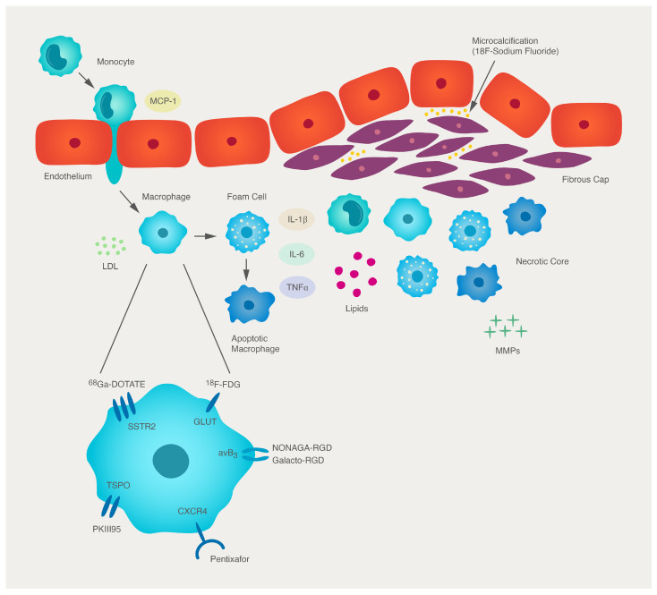
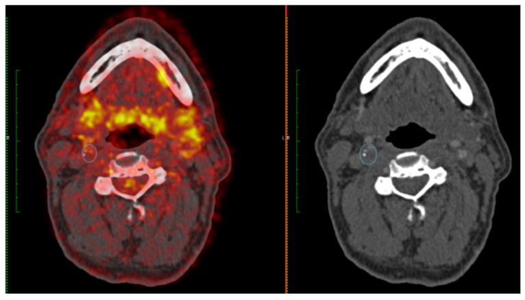
Atherosclerosis is a chronic systemic inflammatory condition of the vasculature and a leading cause of stroke. Luminal stenosis severity is an important factor in determining vascular risk. Conventional imaging modalities, such as angiography or duplex ultrasonography, are used to quantify stenosis severity and inform clinical care but provide limited information on plaque biology. Inflammatory processes are central to atherosclerotic plaque progression and destabilization. 18F-fluorodeoxyglucose (FDG) positron emission tomography (PET) is a validated technique for quantifying plaque inflammation. In this review, we discuss the evolution of FDG-PET as an imaging modality to quantify plaque vulnerability, challenges in standardization of image acquisition and analysis, its potential application to routine clinical care after stroke, and the possible role it will play in future drug discovery.
动脉粥样硬化是一种慢性系统性血管炎症疾病,也是中风的主要病因。管腔狭窄程度是决定血管风险的重要因素。血管造影或双功能超声等传统成像方式用于量化狭窄程度并为临床治疗提供信息,但提供的斑块生物学信息有限。炎症过程是动脉粥样硬化斑块进展和不稳定的核心。18F-氟脱氧葡萄糖(FDG)正电子发射断层扫描(PET)是一种经过验证的技术,可用于定量斑块炎症。在这篇综述中,我们讨论了 FDG-PET 作为一种成像方式来量化斑块易损性的演变、图像采集和分析标准化的挑战、其在中风后常规临床护理中的潜在应用以及它在未来药物发现中可能发挥的作用。