MassGeneral Institute for Neurodegenerative Disease, Massachusetts General Hospital, Harvard Medical School, 114, 16th Street, Boston, MA, 02129, USA.
J. Philip Kistler Stroke Research Center, Massachusetts General Hospital, Harvard Medical School, 175 Cambridge Street, Boston, MA, 02114, USA.
J Neuroinflammation. 2023 Aug 27;20(1):195. doi: 10.1186/s12974-023-02872-0.
Cortical superficial siderosis (cSS) has recently emerged as one of the most important predictors of symptomatic intracerebral hemorrhage and is a risk factor for post-stroke dementia in cerebral amyloid angiopathy (CAA). However, it remains unknown whether cSS is just a marker of severe CAA pathology or may itself contribute to intracerebral hemorrhage risk and cognitive decline. cSS is a chronic manifestation of convexal subarachnoid hemorrhage and is neuropathologically characterized by iron deposits in the superficial cortical layers. We hypothesized that these iron deposits lead to local neuroinflammation, a potentially contributory pathway towards secondary tissue injury.
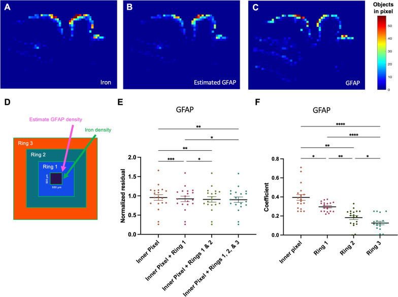
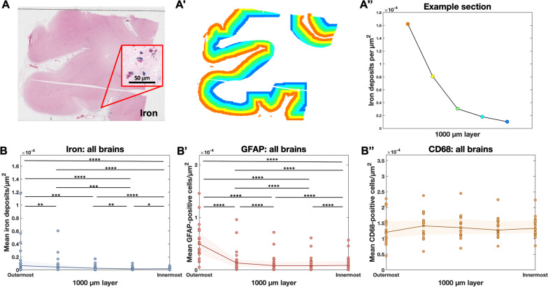
Accordingly, we assessed the distribution of inflammatory markers in relation to cortical iron deposits in post-mortem tissue from CAA cases. Serial sections from the frontal, parietal, temporal, and occipital lobes of nineteen autopsy cases with CAA were stained with Perls' Prussian blue (iron) and underwent immunohistochemistry against glial fibrillary acidic protein (GFAP, reactive astrocytes) and cluster of differentiation 68 (CD68, activated microglia/macrophages). Digitized sections were uploaded to the cloud-based Aiforia platform, where deep-learning algorithms were utilized to detect tissue, iron deposits, and GFAP-positive and CD68-positive cells.
We observed a strong local relationship between cortical iron deposits and reactive astrocytes. Like cSS-related iron, reactive astrocytes were mainly found in the most superficial layers of the cortex. Although we observed iron within both astrocytes and activated microglia/macrophages on co-stains, there was no clear local relationship between the density of microglia/macrophages and the density of iron deposits.
Iron deposition resulting from cSS is associated with local reactive astrogliosis.
皮质表面铁沉积(cSS)最近已成为症状性脑出血的最重要预测因子之一,也是脑淀粉样血管病(CAA)中卒中后痴呆的危险因素。然而,目前尚不清楚 cSS 是否仅是严重 CAA 病理的标志物,或者其本身是否会增加脑出血风险和认知能力下降。cSS 是脑凸面蛛网膜下腔出血的慢性表现,其神经病理学特征为皮质浅层铁沉积。我们假设这些铁沉积会导致局部神经炎症,这可能是导致继发性组织损伤的一个潜在途径。
因此,我们评估了炎症标志物在 CAA 病例死后组织中与皮质铁沉积的分布关系。从 19 例 CAA 尸检病例的额、顶、颞和枕叶连续切片进行普鲁士蓝染色(铁)和胶质纤维酸性蛋白(GFAP,反应性星形胶质细胞)和分化簇 68(CD68,活化的小胶质细胞/巨噬细胞)免疫组化染色。数字化切片上传至基于云的 Aiforia 平台,该平台利用深度学习算法检测组织、铁沉积、GFAP 阳性和 CD68 阳性细胞。
我们观察到皮质铁沉积与反应性星形胶质细胞之间存在很强的局部关系。与 cSS 相关的铁一样,反应性星形胶质细胞主要存在于皮质的最浅层。虽然我们在共染色时观察到铁存在于星形胶质细胞和活化的小胶质细胞/巨噬细胞内,但小胶质细胞/巨噬细胞密度与铁沉积密度之间没有明确的局部关系。
cSS 引起的铁沉积与局部反应性星形胶质增生有关。