Department of Neurosurgery, LMU University Hospital, LMU Munich, Marchioninistrasse 15, 81377, Munich, Germany.
German Cancer Consortium (DKTK), Partner Site Munich, Munich, Germany.
Eur J Nucl Med Mol Imaging. 2023 Dec;51(1):206-217. doi: 10.1007/s00259-023-06400-3. Epub 2023 Aug 29.
Tumor resection represents the first-line treatment for symptomatic meningiomas, and the extent of resection has been shown to be of prognostic importance. Assessment of tumor remnants with somatostatin receptor PET proves to be superior to intraoperative estimation with Simpson grading or MRI. In this preliminary study, we evaluate the prognostic relevance of postoperative PET for progression-free survival in meningiomas.
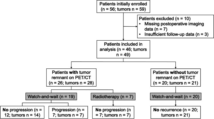
We conducted a post hoc analysis on a prospective patient cohort with resected meningioma WHO grade 1. Patients received postoperative MRI and [Ga]Ga-DOTA-TATE PET/CT and were followed regularly with MRI surveillance scans for detection of tumor recurrence/progression.
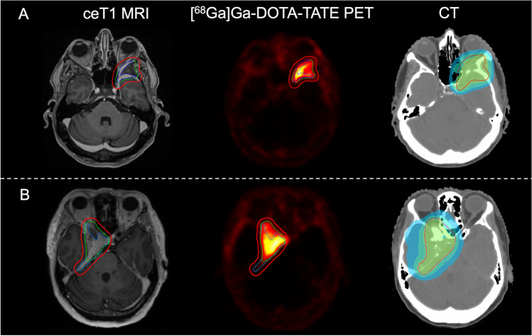
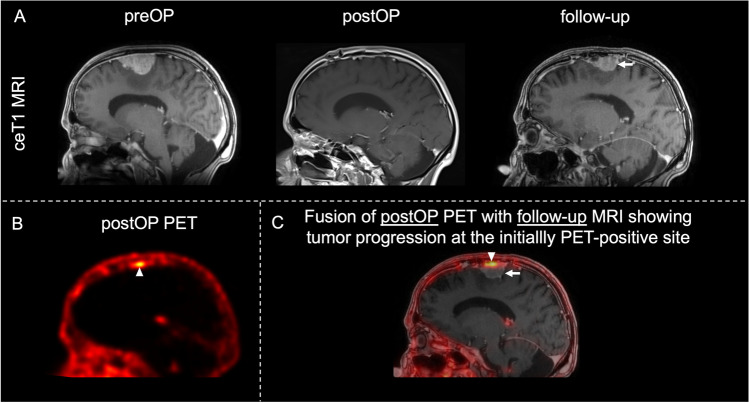
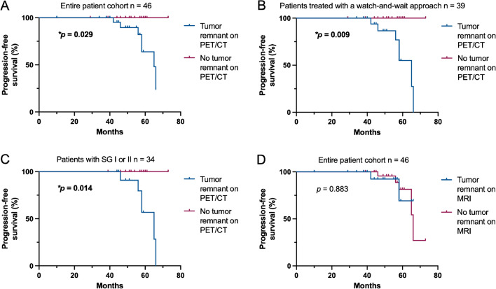
We included 46 patients with 49 tumors. The mean age at diagnosis was 57.8 ± 1.7 years with a male-to-female ratio of 1:1.7. Local tumor progression occurred in 7/49 patients (14%) after a median follow-up of 52 months. Positive PET was associated with an increased risk for progression (*p = 0.015) and a lower progression-free survival (*p = 0.029), whereas MRI was not. 20 out of 20 patients (100%) with negative PET findings remained recurrence-free. The location of recurrence/progression on MRI was adjacent to regions where postoperative PET indicated tumor remnants in all cases. Gross tumor volumes were higher on PET compared to MRI (*p = 0.032).
Our data show that [Ga]Ga-DOTA-TATE PET/CT is highly sensitive in revealing tumor remnants in patients with meningioma WHO grade 1. Negative PET findings were associated with a higher progression-free survival, thus improving surveillance. In patients with tumor remnants, additional PET can optimize adjuvant radiotherapy target planning of surgically resected meningiomas.
肿瘤切除术是治疗有症状脑膜瘤的一线治疗方法,且已证实切除范围与预后有关。使用生长抑素受体 PET 评估肿瘤残余物优于术中辛普森分级或 MRI 评估。在这项初步研究中,我们评估了脑膜瘤术后 PET 对无进展生存期的预后相关性。
我们对接受手术切除的 WHO 分级 1 级脑膜瘤的前瞻性患者队列进行了事后分析。患者术后接受 MRI 和 [Ga]Ga-DOTA-TATE PET/CT 检查,并定期进行 MRI 随访扫描以检测肿瘤复发/进展。
我们纳入了 46 例 49 个肿瘤患者。诊断时的平均年龄为 57.8±1.7 岁,男女比例为 1:1.7。49 例肿瘤中有 7 例(14%)在中位随访 52 个月后出现局部肿瘤进展。阳性 PET 与进展风险增加相关(*p=0.015),无进展生存期降低(*p=0.029),而 MRI 无此相关性。20 例 PET 结果阴性的患者(100%)均未复发。MRI 上的复发/进展部位与术后 PET 显示肿瘤残余的部位相邻,所有病例均如此。与 MRI 相比,PET 上的肿瘤总体积更高(*p=0.032)。
我们的数据表明,[Ga]Ga-DOTA-TATE PET/CT 对揭示 WHO 分级 1 级脑膜瘤患者的肿瘤残余非常敏感。阴性 PET 结果与更高的无进展生存期相关,从而改善了监测。对于有肿瘤残余的患者,额外的 PET 可优化手术切除脑膜瘤的辅助放疗靶区规划。