Department of Medicine II, Heart Center, University Hospital Bonn, Bonn, Germany.
Department of Cardiology and Internal Intensive Care, Asklepios Clinic St. Georg, Hamburg, Germany.
Sci Rep. 2023 Aug 30;13(1):14181. doi: 10.1038/s41598-023-41054-z.
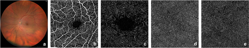
Cerebral embolization is a known complication of transcatheter aortic valve implantation (TAVI) but the effect of the procedure on the ocular perfusion is currently unclear. Thus, we investigated post-procedural morphologic and perfusion changes of the retina and choroid, using optical coherence tomography angiography (OCTA) and color fundus photography (CFP) in a prospective cohort study. Ophthalmic examinations were conducted pre- and post-TAVI. OCTA images were analyzed quantitatively based on vessel density and skeleton density of the superficial and deep retinal plexus as well as the signal intensity and flow deficits in the choriocapillaris. CFP images were assessed for presence of acute retinal ischemia, optic nerve swelling, vessel emboli, hemorrhages and cotton wool spots. Data was analyzed using linear mixed models. Twenty patients (9 women; 11 men) at a mean age of 81 ± 6 years were included. Pre- and post-interventional ocular imaging data were available for 32 eyes. The analysis revealed a significant impairment of the choriocapillaris perfusion after TAVI with an increased proportion of flow deficits (p = 0.044). When controlling for blood pressure, the average size of choriocapillaris flow voids was significantly increased (systolic and diastolic, p = 0.039 and 0.029). Qualitatively, focal areas of retinal ischemia were detected on OCTA in 33% of participants. Silent emboli or cotton wool spots were identified on CFP in 21%. Our findings indicate a reduced choroidal perfusion as well as areas of retinal ischemia and embolization in a considerable proportion of patients following TAVI. Pending confirmation in a larger sample, these complications merit monitoring as well as inclusion in consent procedures for TAVI.
经导管主动脉瓣植入术(TAVI)后发生脑栓塞是已知的并发症,但该手术对眼灌注的影响目前尚不清楚。因此,我们通过前瞻性队列研究,使用光学相干断层扫描血管造影(OCTA)和眼底彩色照相(CFP)检查,研究视网膜和脉络膜的术后形态和灌注变化。在 TAVI 前后进行眼科检查。根据浅层和深层视网膜丛的血管密度和骨架密度以及脉络膜毛细血管的信号强度和血流缺损,对 OCTA 图像进行定量分析。对 CFP 图像进行评估,以发现急性视网膜缺血、视神经肿胀、血管栓塞、出血和棉絮斑。使用线性混合模型进行数据分析。20 名患者(9 名女性;11 名男性),平均年龄 81±6 岁,纳入研究。共有 32 只眼获得了术前和术后的眼部影像学数据。分析显示,TAVI 后脉络膜毛细血管灌注明显受损,血流缺损比例增加(p=0.044)。当控制血压时,脉络膜毛细血管血流缺失的平均大小显著增加(收缩压和舒张压,p=0.039 和 0.029)。定性分析发现,33%的参与者在 OCTA 上检测到局部视网膜缺血。21%的参与者在 CFP 上发现有隐形栓塞或棉絮斑。我们的研究结果表明,TAVI 后相当一部分患者的脉络膜灌注减少,以及视网膜缺血和栓塞区。在更大的样本中得到证实之前,这些并发症值得监测,并纳入 TAVI 的同意程序。