Department of Ultrasound, The Third Xiangya Hospital, Central South University, Changsha, 410013, Hunan, China.
BMC Cancer. 2023 Aug 30;23(1):813. doi: 10.1186/s12885-023-11336-w.
Lymphovascular invasion (LVI) indicates resistance to preoperative adjuvant chemotherapy and a poor prognosis and can only be diagnosed by postoperative pathological examinations in breast cancer. Thus, a technique for preoperative diagnosis of LVI is urgently needed. We aim to explore the ability of an automated breast volume scanner (ABVS)-based radiomics model to noninvasively predict the LVI status in breast cancer.
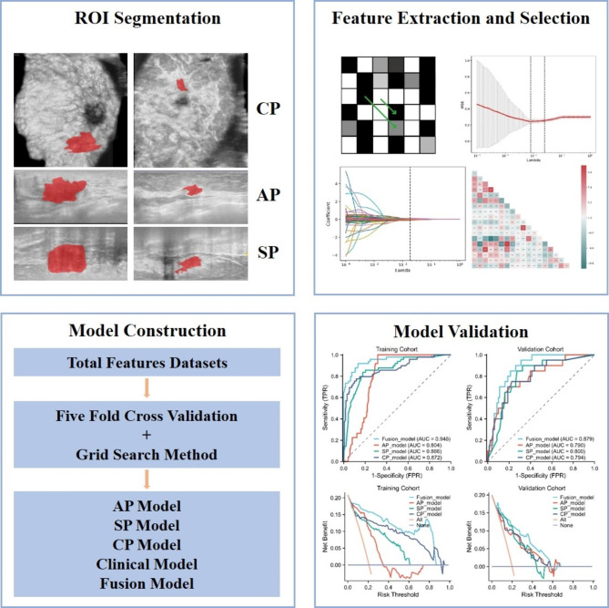
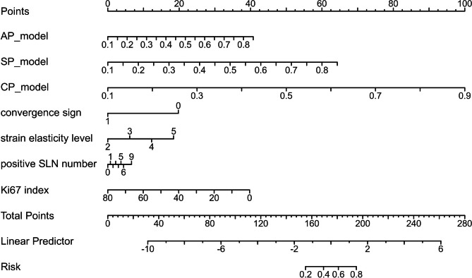
We conducted a retrospective analysis of data from 335 patients diagnosed with T1-3 breast cancer between October 2019 and September 2022. The patients were divided into training cohort and validation cohort with a ratio of 7:3. For each patient, 5901 radiomics features were extracted from ABVS images. Feature selection was performed using LASSO method. We created machine learning models for different feature sets with support vector machine algorithm to predict LVI. And significant clinicopathologic factors were identified by univariate and multivariate logistic regression to combine with three radiomics signatures as to develop a fusion model.
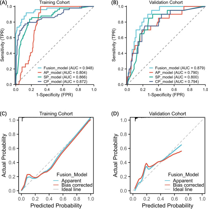
The three SVM-based prediction models, demonstrated relatively high efficacy in identifying LVI of breast cancer, with AUCs of 79.00%, 80.00% and 79.40% and an accuracy of 71.00%, 80.00% and 75.00% in the validation cohort for AP, SP and CP plane image. The fusion model achieved the highest AUC of 87.90% and an accuracy of 85.00% in the validation cohort.
The combination of radiomics features from ABVS images and an SVM prediction model showed promising performance for preoperative noninvasive prediction of LVI in breast cancer.
淋巴管浸润(LVI)表明对术前辅助化疗有抵抗力和预后不良,只能通过乳腺癌术后病理检查来诊断。因此,迫切需要一种术前诊断 LVI 的技术。我们旨在探讨基于自动乳腺容积扫描仪(ABVS)的放射组学模型无创预测乳腺癌 LVI 状态的能力。
我们对 2019 年 10 月至 2022 年 9 月期间诊断为 T1-3 期乳腺癌的 335 例患者的数据进行了回顾性分析。患者分为训练队列和验证队列,比例为 7:3。对于每位患者,从 ABVS 图像中提取 5901 个放射组学特征。使用 LASSO 方法进行特征选择。我们使用支持向量机算法为不同的特征集创建机器学习模型,以预测 LVI。并通过单因素和多因素逻辑回归确定显著的临床病理因素,将其与三个放射组学特征相结合,建立融合模型。
三种基于 SVM 的预测模型在验证队列中识别乳腺癌 LVI 的效能较高,AP、SP 和 CP 平面图像的 AUC 分别为 79.00%、80.00%和 79.40%,准确性分别为 71.00%、80.00%和 75.00%。融合模型在验证队列中获得了最高的 AUC 为 87.90%和准确性为 85.00%。
ABVS 图像的放射组学特征与 SVM 预测模型的结合,为乳腺癌术前无创预测 LVI 提供了有前途的方法。