• 文献检索
  • 文档翻译
  • 深度研究
  • 学术资讯
  • Suppr Zotero 插件Zotero 插件
  • 邀请有礼
  • 套餐&价格
  • 历史记录
应用&插件
Suppr Zotero 插件Zotero 插件浏览器插件Mac 客户端Windows 客户端微信小程序
定价
高级版会员购买积分包购买API积分包
服务
文献检索文档翻译深度研究API 文档MCP 服务
关于我们
关于 Suppr公司介绍联系我们用户协议隐私条款
关注我们

Suppr 超能文献

核心技术专利:CN118964589B侵权必究
粤ICP备2023148730 号-1Suppr @ 2026

文献检索

告别复杂PubMed语法,用中文像聊天一样搜索,搜遍4000万医学文献。AI智能推荐,让科研检索更轻松。

立即免费搜索

文件翻译

保留排版,准确专业,支持PDF/Word/PPT等文件格式,支持 12+语言互译。

免费翻译文档

深度研究

AI帮你快速写综述,25分钟生成高质量综述,智能提取关键信息,辅助科研写作。

立即免费体验

特定扫描仪CT模拟器的开发、验证与简化

Development, validation, and simplification of a scanner-specific CT simulator.

作者信息

Tunissen Sjoerd A M, Oostveen Luuk J, Moriakov Nikita, Teuwen Jonas, Michielsen Koen, Smit Ewoud J, Sechopoulos Ioannis

机构信息

Department of Medical Imaging, Radboudumc, Nijmegen, The Netherlands.

Department of Radiation Oncology, Netherlands Cancer Institute, Amsterdam, The Netherlands.

出版信息

Med Phys. 2024 Mar;51(3):2081-2095. doi: 10.1002/mp.16679. Epub 2023 Sep 1.

DOI:10.1002/mp.16679
PMID:37656009
原文链接:https://pmc.ncbi.nlm.nih.gov/articles/PMC10904672/
Abstract

BACKGROUND

Simulated computed tomography (CT) images allow for knowledge of the underlying ground truth and for easy variation of imaging conditions, making them ideal for testing and optimization of new applications or algorithms. However, simulating all processes that affect CT images can result in simulations that are demanding in terms of processing time and computer memory. Therefore, it is of interest to determine how much the simulation can be simplified while still achieving realistic results.

PURPOSE

To develop a scanner-specific CT simulation using physics-based simulations for the position-dependent effects and shift-invariant image corruption methods for the detector effects. And to investigate the impact on image realism of introducing simplifications in the simulation process that lead to faster and less memory-demanding simulations.

METHODS

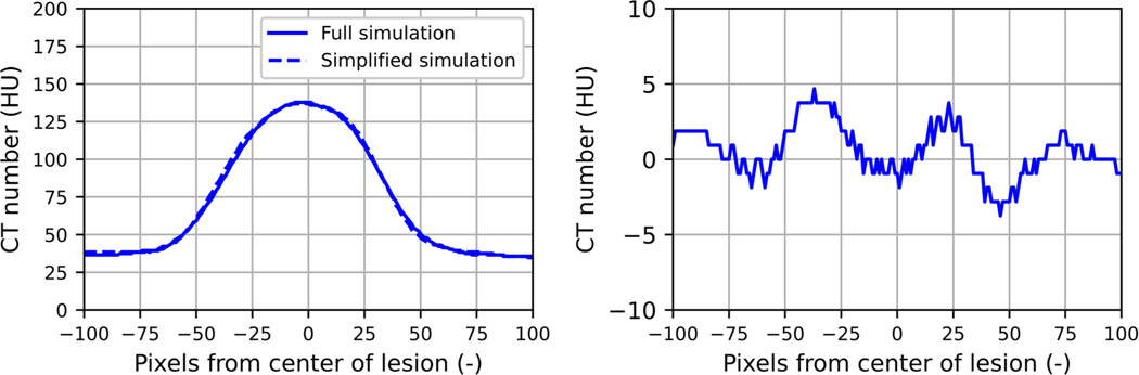
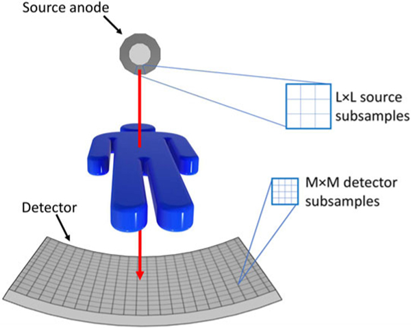
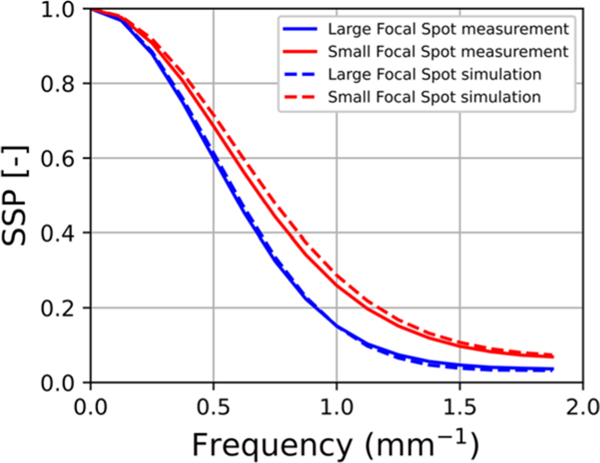
To make the simulator realistic and scanner-specific, the spatial resolution and noise characteristics, and the exposure-to-detector output relationship of a clinical CT system were determined. The simulator includes a finite focal spot size, raytracing of the digital phantom, gantry rotation during projection acquisition, and finite detector element size. Previously published spectral models were used to model the spectrum for the given tube voltage. The integrated energy at each element of the detector was calculated using the Beer-Lambert law. The resulting angular projections were subsequently corrupted by the detector modulation transfer function (MTF), and by addition of noise according to the noise power spectrum (NPS) and signal mean-variance relationship, which were measured for different scanner settings. The simulated sinograms were reconstructed on the clinical CT system and compared to real CT images in terms of CT numbers, noise magnitude using the standard deviation, noise frequency content using the NPS, and spatial resolution using the MTF throughout the field of view (FOV). The CT numbers were validated using a multi-energy CT phantom, the noise magnitude and frequency were validated with a water phantom, and the spatial resolution was validated with a tungsten wire. These metrics were compared at multiple scanner settings, and locations in the FOV. Once validated, the simulation was simplified by reducing the level of subsampling of the focal spot area, rotation and of detector pixel size, and the changes in MTFs were analyzed.

RESULTS

The average relative errors for spatial resolution within and across image slices, noise magnitude, and noise frequency content within and across slices were 3.4%, 3.3%, 4.9%, 3.9%, and 6.2%, respectively. The average absolute difference in CT numbers was 10.2 HU and the maximum was 22.5 HU. The simulation simplification showed that all subsampling can be avoided, except for angular, while the error in frequency at 10% MTF would be maximum 16.3%.

CONCLUSION

The simulation of a scanner-specific CT allows for the generation of realistic CT images by combining physics-based simulations for the position-dependent effects and image-corruption methods for the shift-invariant ones. Together with the available ground truth of the digital phantom, it results in a useful tool to perform quantitative analysis of reconstruction or post-processing algorithms. Some simulation simplifications allow for reduced time and computer power requirements with minimal loss of realism.

摘要

背景

模拟计算机断层扫描(CT)图像有助于了解潜在的真实情况,并便于改变成像条件,使其成为测试和优化新应用或算法的理想选择。然而,模拟所有影响CT图像的过程可能会导致模拟在处理时间和计算机内存方面要求很高。因此,确定在仍能获得逼真结果的同时,模拟可以简化到何种程度是很有意义的。

目的

开发一种特定于扫描仪的CT模拟,使用基于物理的模拟来处理位置相关效应,并使用平移不变图像损坏方法来处理探测器效应。并研究在模拟过程中引入简化措施对图像逼真度的影响,这些简化措施可实现更快且对内存要求更低的模拟。

方法

为使模拟器逼真且特定于扫描仪,确定了临床CT系统的空间分辨率、噪声特性以及曝光与探测器输出的关系。该模拟器包括有限的焦点尺寸、数字体模的光线追踪、投影采集期间的机架旋转以及有限的探测器元件尺寸。使用先前发表的光谱模型对给定管电压下的光谱进行建模。利用比尔-朗伯定律计算探测器每个元件处的积分能量。随后,所得的角度投影通过探测器调制传递函数(MTF)进行损坏,并根据噪声功率谱(NPS)和信号均值-方差关系添加噪声,这些关系是针对不同扫描仪设置测量得到的。在临床CT系统上重建模拟的正弦图,并在整个视野(FOV)内,就CT值、使用标准差表示的噪声幅度、使用NPS表示的噪声频率内容以及使用MTF表示的空间分辨率而言,将其与真实CT图像进行比较。使用多能量CT体模验证CT值,使用水体模验证噪声幅度和频率,使用钨丝验证空间分辨率。在多个扫描仪设置以及FOV中的不同位置比较这些指标。一旦验证完成,通过降低焦点区域子采样级别、旋转以及探测器像素尺寸来简化模拟,并分析MTF的变化。

结果

图像切片内和跨切片的空间分辨率、噪声幅度以及噪声频率内容的平均相对误差分别为3.4%、3.3%、4.9%、3.9%和6.2%。CT值的平均绝对差值为10.2 HU,最大值为22.5 HU。模拟简化表明,除角度外所有子采样均可避免,而在MTF为10%时频率误差最大为16.3%。

结论

特定于扫描仪的CT模拟通过结合基于物理的模拟来处理位置相关效应以及针对平移不变效应的图像损坏方法,能够生成逼真的CT图像。连同数字体模的可用真实情况,它成为对重建或后处理算法进行定量分析的有用工具。一些模拟简化措施可在对逼真度影响最小的情况下降低时间和计算机功率要求。

https://cdn.ncbi.nlm.nih.gov/pmc/blobs/a06b/10904672/cfca3086d28c/nihms-1939930-f0017.jpg
https://cdn.ncbi.nlm.nih.gov/pmc/blobs/a06b/10904672/6d5fdf0d54a5/nihms-1939930-f0001.jpg
https://cdn.ncbi.nlm.nih.gov/pmc/blobs/a06b/10904672/5b7bd9c9f11f/nihms-1939930-f0002.jpg
https://cdn.ncbi.nlm.nih.gov/pmc/blobs/a06b/10904672/021f5d5abed4/nihms-1939930-f0003.jpg
https://cdn.ncbi.nlm.nih.gov/pmc/blobs/a06b/10904672/1ff34882ffc9/nihms-1939930-f0004.jpg
https://cdn.ncbi.nlm.nih.gov/pmc/blobs/a06b/10904672/d36a4d268c04/nihms-1939930-f0005.jpg
https://cdn.ncbi.nlm.nih.gov/pmc/blobs/a06b/10904672/7c205b972ce1/nihms-1939930-f0006.jpg
https://cdn.ncbi.nlm.nih.gov/pmc/blobs/a06b/10904672/c49e54d07e21/nihms-1939930-f0007.jpg
https://cdn.ncbi.nlm.nih.gov/pmc/blobs/a06b/10904672/74495428421f/nihms-1939930-f0008.jpg
https://cdn.ncbi.nlm.nih.gov/pmc/blobs/a06b/10904672/cb8bbc5edeeb/nihms-1939930-f0009.jpg
https://cdn.ncbi.nlm.nih.gov/pmc/blobs/a06b/10904672/11aaccbb7591/nihms-1939930-f0010.jpg
https://cdn.ncbi.nlm.nih.gov/pmc/blobs/a06b/10904672/754eaacf5efc/nihms-1939930-f0011.jpg
https://cdn.ncbi.nlm.nih.gov/pmc/blobs/a06b/10904672/44b798852087/nihms-1939930-f0012.jpg
https://cdn.ncbi.nlm.nih.gov/pmc/blobs/a06b/10904672/43b943d5e8f6/nihms-1939930-f0013.jpg
https://cdn.ncbi.nlm.nih.gov/pmc/blobs/a06b/10904672/431a73b0d722/nihms-1939930-f0014.jpg
https://cdn.ncbi.nlm.nih.gov/pmc/blobs/a06b/10904672/7917b1c4c64b/nihms-1939930-f0015.jpg
https://cdn.ncbi.nlm.nih.gov/pmc/blobs/a06b/10904672/8614894d7540/nihms-1939930-f0016.jpg
https://cdn.ncbi.nlm.nih.gov/pmc/blobs/a06b/10904672/cfca3086d28c/nihms-1939930-f0017.jpg

相似文献

1
Development, validation, and simplification of a scanner-specific CT simulator.特定扫描仪CT模拟器的开发、验证与简化
Med Phys. 2024 Mar;51(3):2081-2095. doi: 10.1002/mp.16679. Epub 2023 Sep 1.
2
Validation of synthesized normal-resolution image data generated from high-resolution acquisitions on a commercial CT scanner.在商用CT扫描仪上对由高分辨率采集生成的合成正常分辨率图像数据进行验证。
Med Phys. 2020 Oct;47(10):4775-4785. doi: 10.1002/mp.14395. Epub 2020 Aug 5.
3
Voxelwise characterization of noise for a clinical photon-counting CT scanner with a model-based iterative reconstruction algorithm.基于模型的迭代重建算法的临床光子计数CT扫描仪噪声的体素特征分析。
Eur Radiol Exp. 2025 Jan 2;9(1):2. doi: 10.1186/s41747-024-00541-2.
4
DukeSim: A Realistic, Rapid, and Scanner-Specific Simulation Framework in Computed Tomography.杜克仿真:计算机断层扫描中的一种真实、快速且与扫描仪特定的仿真框架。
IEEE Trans Med Imaging. 2019 Jun;38(6):1457-1465. doi: 10.1109/TMI.2018.2886530. Epub 2018 Dec 12.
5
Development and tuning of models for accurate simulation of CT spatial resolution using CatSim.使用CatSim开发和调整用于精确模拟CT空间分辨率的模型。
Phys Med Biol. 2024 Feb 8;69(4). doi: 10.1088/1361-6560/ad2122.
6
Effects of ray profile modeling on resolution recovery in clinical CT.射线轮廓建模对临床 CT 中分辨率恢复的影响。
Med Phys. 2014 Feb;41(2):021907. doi: 10.1118/1.4862510.
7
Realistic simulation of reduced-dose CT with noise modeling and sinogram synthesis using DICOM CT images.使用DICOM CT图像通过噪声建模和正弦图合成对低剂量CT进行逼真模拟。
Med Phys. 2014 Jan;41(1):011901. doi: 10.1118/1.4830431.
8
Characteristics and clinical application of a treatment simulator with Ct-option.具有CT选项的治疗模拟器的特性与临床应用
Radiother Oncol. 1999 Mar;50(3):355-66. doi: 10.1016/s0167-8140(99)00033-x.
9
Towards 4D dedicated breast CT perfusion imaging of cancer: development and validation of computer simulated images.面向癌症的 4D 专用乳腺 CT 灌注成像:计算机模拟图像的开发和验证。
Phys Med Biol. 2019 Dec 13;64(24):245004. doi: 10.1088/1361-6560/ab55ac.
10
Exploration of the pulse pileup effects in a clinical CdTe-based photon-counting computed tomography.临床基于 CdTe 的光子计数 CT 中的脉搏堆积效应的探索。
Med Phys. 2023 Nov;50(11):6693-6703. doi: 10.1002/mp.16671. Epub 2023 Aug 21.

引用本文的文献

1
Simulating scanner-and algorithm-specific 3D CT noise texture using physics-informed 2D and 2.5D generative neural network models.使用物理信息二维和2.5维生成神经网络模型模拟特定扫描仪和算法的三维CT噪声纹理
Proc SPIE Int Soc Opt Eng. 2025 Feb;13405. doi: 10.1117/12.3047909. Epub 2025 Apr 8.
2
Physics-informed model-based generative neural network for synthesizing scanner- and algorithm-specific low-dose CT exams.基于物理信息模型的生成式神经网络,用于合成特定扫描仪和算法的低剂量CT检查。
Med Phys. 2025 Jun;52(6):3940-3958. doi: 10.1002/mp.17680. Epub 2025 Feb 13.
3
Performance evaluation of a 4D similarity filter for dynamic CT angiography imaging of the liver.

本文引用的文献

1
XCIST-an open access x-ray/CT simulation toolkit.XCIST-一个开放获取的 X 射线/CT 模拟工具包。
Phys Med Biol. 2022 Sep 28;67(19). doi: 10.1088/1361-6560/ac9174.
2
Development of a computational phantom for validation of automated noise measurement in CT images.用于验证 CT 图像中自动噪声测量的计算体模的开发。
Biomed Phys Eng Express. 2020 Sep 29;6(6). doi: 10.1088/2057-1976/abb2f8.
3
Virtual clinical trials in medical imaging: a review.医学成像中的虚拟临床试验:综述
用于肝脏动态CT血管造影成像的4D相似性滤波器的性能评估
Med Phys. 2024 Dec;51(12):8814-8827. doi: 10.1002/mp.17394. Epub 2024 Sep 12.
J Med Imaging (Bellingham). 2020 Jul;7(4):042805. doi: 10.1117/1.JMI.7.4.042805. Epub 2020 Apr 11.
4
Reduced Lung-Cancer Mortality with Volume CT Screening in a Randomized Trial.随机试验中 CT 容积筛查降低肺癌死亡率
N Engl J Med. 2020 Feb 6;382(6):503-513. doi: 10.1056/NEJMoa1911793. Epub 2020 Jan 29.
5
Towards 4D dedicated breast CT perfusion imaging of cancer: development and validation of computer simulated images.面向癌症的 4D 专用乳腺 CT 灌注成像:计算机模拟图像的开发和验证。
Phys Med Biol. 2019 Dec 13;64(24):245004. doi: 10.1088/1361-6560/ab55ac.
6
Trends in Use of Medical Imaging in US Health Care Systems and in Ontario, Canada, 2000-2016.2000-2016 年美国医疗保健系统和加拿大安大略省医疗成像使用趋势。
JAMA. 2019 Sep 3;322(9):843-856. doi: 10.1001/jama.2019.11456.
7
Technical Note: PYRO-NN: Python reconstruction operators in neural networks.技术说明:PYRO-NN:神经网络中的 Python 重建算子。
Med Phys. 2019 Nov;46(11):5110-5115. doi: 10.1002/mp.13753. Epub 2019 Aug 27.
8
DukeSim: A Realistic, Rapid, and Scanner-Specific Simulation Framework in Computed Tomography.杜克仿真:计算机断层扫描中的一种真实、快速且与扫描仪特定的仿真框架。
IEEE Trans Med Imaging. 2019 Jun;38(6):1457-1465. doi: 10.1109/TMI.2018.2886530. Epub 2018 Dec 12.
9
Deep Convolutional Framelet Denosing for Low-Dose CT via Wavelet Residual Network.基于小波残差网络的低剂量 CT 深度学习卷积框架去噪。
IEEE Trans Med Imaging. 2018 Jun;37(6):1358-1369. doi: 10.1109/TMI.2018.2823756.
10
A deep convolutional neural network using directional wavelets for low-dose X-ray CT reconstruction.一种基于方向小波的深度卷积神经网络在低剂量 X 射线 CT 重建中的应用。
Med Phys. 2017 Oct;44(10):e360-e375. doi: 10.1002/mp.12344.