Departments of Pediatrics & Biochemistry and Molecular Biology, Dalhousie University, Halifax, NS, Canada.
Department of Pathology, Dalhousie University, Halifax, NS, Canada.
Cell Death Dis. 2023 Sep 1;14(9):580. doi: 10.1038/s41419-023-06106-2.
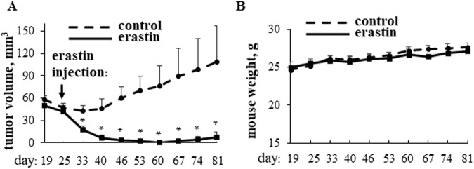
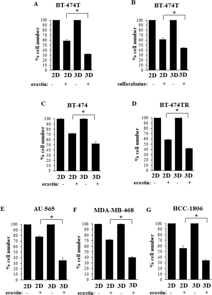
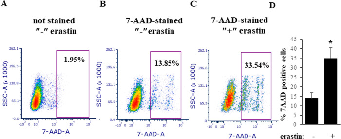
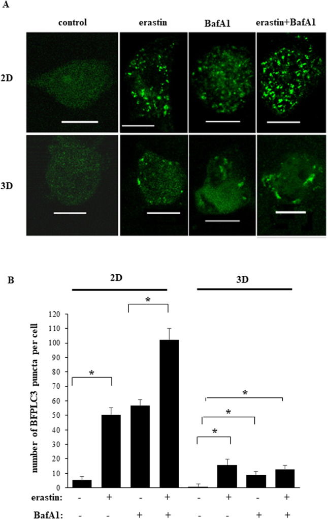
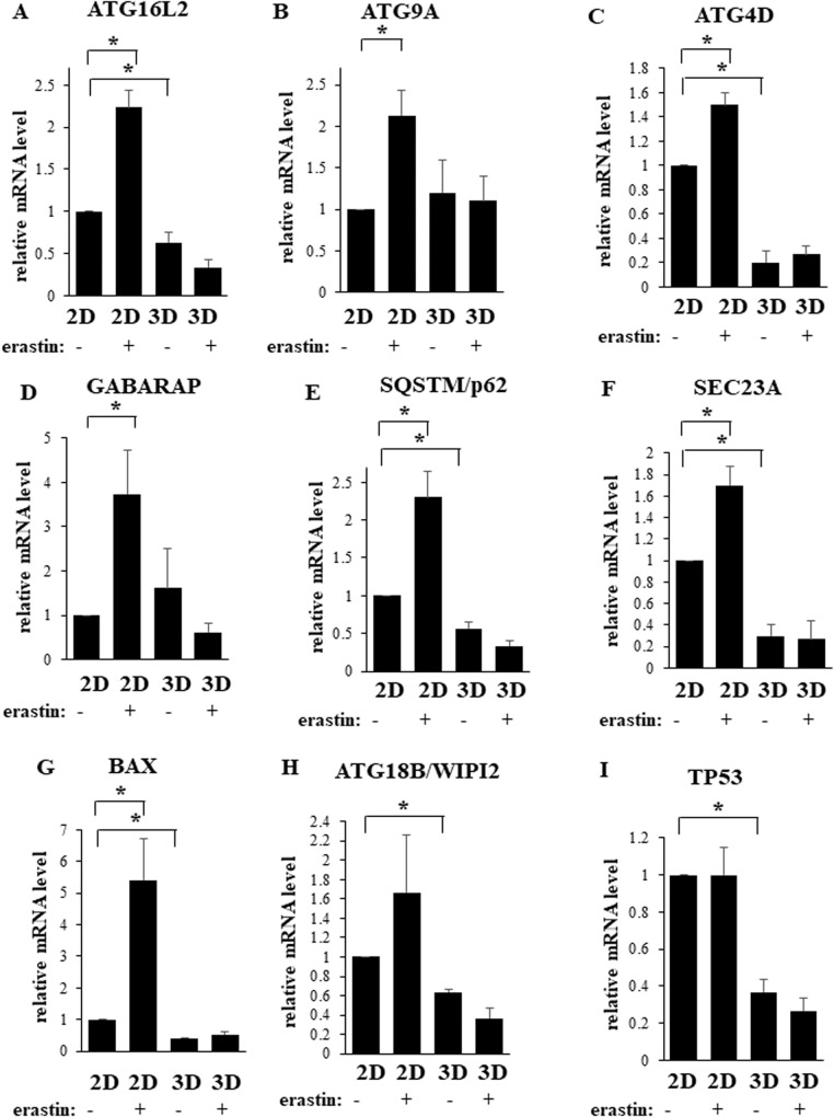
Drugs causing ferroptosis, iron-mediated cell death, represent promising tools for cancer treatment. While exploring the effect of these drugs on breast cancer (BC), we found that a ferroptosis-inducing drug erastin dramatically inhibits tumorigenicity of human BC cells in mice but when used at a concentration known to effectively kill other cell types only modestly reduces such growth in 2D monolayer culture. BCs grow in vivo as 3D masses, and we found that ferroptosis inducers erastin and sulfasalazine inhibit growth of multiple human BC cell lines in 3D culture significantly stronger than in 2D culture. To understand the mechanism of this differential effect, we found that ferroptosis inducers upregulate mRNAs encoding multiple direct and indirect autophagy stimulators, such as ATG16L2, ATG9A, ATG4D, GABARAP, SQSTM/p62, SEC23A and BAX, in tumor cells growing in 2D but not in 3D culture. Furthermore, these drugs promoted autophagy of tumor cells growing in a 2D but not in a 3D manner. We observed that pharmacological inhibition of autophagy-stimulating protein kinase ULK1 or RNA interference-mediated knockdown of autophagy mediator ATG12 significantly sensitized tumor cells to erastin treatment in 2D culture. We also found that ferroptosis-promoting treatments upregulate heme oxygenase-1 (HO-1) in BC cells. HO-1 increases cellular free iron pool and can potentially promote ferroptosis. Indeed, we observed that HO-1 knockdown by RNA interference reversed the effect of ferroptosis inducers on BC cell 3D growth. Hence, the effect of these drugs on such growth is mediated by HO-1. In summary, autophagy triggered by ferroptosis-promoting drugs reduces their ability to kill BC growing in a 2D manner. This protection mechanism is inhibited in BC cells growing as a 3D mass, and ferroptosis-promoting drugs kill such cells more effectively. Moreover, this death is mediated by HO-1. Thus, ferroptosis induction represents a promising strategy for blocking 3D BC growth.
导致铁死亡的药物,即铁介导的细胞死亡,代表了癌症治疗的有前途的工具。在探索这些药物对乳腺癌(BC)的影响时,我们发现铁死亡诱导剂 erastin 可显著抑制小鼠中人类 BC 细胞的致瘤性,但在已知可有效杀死其他细胞类型的浓度下,其对 2D 单层培养中的这种生长的抑制作用仅适度。BC 在体内生长为 3D 团块,我们发现铁死亡诱导剂 erastin 和柳氮磺胺吡啶在 3D 培养中显著抑制多种人类 BC 细胞系的生长,其抑制作用强于 2D 培养。为了理解这种差异效应的机制,我们发现铁死亡诱导剂上调了编码多种直接和间接自噬刺激物的 mRNA,如 ATG16L2、ATG9A、ATG4D、GABARAP、SQSTM/p62、SEC23A 和 BAX,在 2D 培养中生长的肿瘤细胞中,但不在 3D 培养中。此外,这些药物促进了在 2D 而非 3D 中生长的肿瘤细胞的自噬。我们观察到,自噬刺激蛋白激酶 ULK1 的药理学抑制或自噬介体 ATG12 的 RNAi 介导的敲低显著增强了肿瘤细胞对 erastin 在 2D 培养中的敏感性。我们还发现,铁死亡促进剂上调了 BC 细胞中的血红素加氧酶-1(HO-1)。HO-1 增加细胞内游离铁池,并可能促进铁死亡。事实上,我们观察到 RNAi 介导的 HO-1 敲低逆转了铁死亡诱导剂对 BC 细胞 3D 生长的影响。因此,这些药物对这种生长的作用是由 HO-1 介导的。总之,铁死亡促进剂触发的自噬降低了它们在 2D 方式下杀死 BC 的能力。这种保护机制在作为 3D 团块生长的 BC 细胞中被抑制,铁死亡促进剂更有效地杀死这些细胞。此外,这种死亡是由 HO-1 介导的。因此,铁死亡诱导代表了阻断 3D BC 生长的有前途的策略。