• 文献检索
  • 文档翻译
  • 深度研究
  • 学术资讯
  • Suppr Zotero 插件Zotero 插件
  • 邀请有礼
  • 套餐&价格
  • 历史记录
应用&插件
Suppr Zotero 插件Zotero 插件浏览器插件Mac 客户端Windows 客户端微信小程序
定价
高级版会员购买积分包购买API积分包
服务
文献检索文档翻译深度研究API 文档MCP 服务
关于我们
关于 Suppr公司介绍联系我们用户协议隐私条款
关注我们

Suppr 超能文献

核心技术专利:CN118964589B侵权必究
粤ICP备2023148730 号-1Suppr @ 2026

文献检索

告别复杂PubMed语法,用中文像聊天一样搜索,搜遍4000万医学文献。AI智能推荐,让科研检索更轻松。

立即免费搜索

文件翻译

保留排版,准确专业,支持PDF/Word/PPT等文件格式,支持 12+语言互译。

免费翻译文档

深度研究

AI帮你快速写综述,25分钟生成高质量综述,智能提取关键信息,辅助科研写作。

立即免费体验

在临床研究环境中整合人工智能工具:卵巢癌应用案例

Integrating Artificial Intelligence Tools in the Clinical Research Setting: The Ovarian Cancer Use Case.

作者信息

Escudero Sanchez Lorena, Buddenkotte Thomas, Al Sa'd Mohammad, McCague Cathal, Darcy James, Rundo Leonardo, Samoshkin Alex, Graves Martin J, Hollamby Victoria, Browne Paul, Crispin-Ortuzar Mireia, Woitek Ramona, Sala Evis, Schönlieb Carola-Bibiane, Doran Simon J, Öktem Ozan

机构信息

Department of Radiology, University of Cambridge, Cambridge CB2 0QQ, UK.

Cancer Research UK Cambridge Centre, Li Ka Shing Centre, Cambridge CB2 0RE, UK.

出版信息

Diagnostics (Basel). 2023 Aug 30;13(17):2813. doi: 10.3390/diagnostics13172813.

DOI:10.3390/diagnostics13172813
PMID:37685352
原文链接:https://pmc.ncbi.nlm.nih.gov/articles/PMC10486639/
Abstract

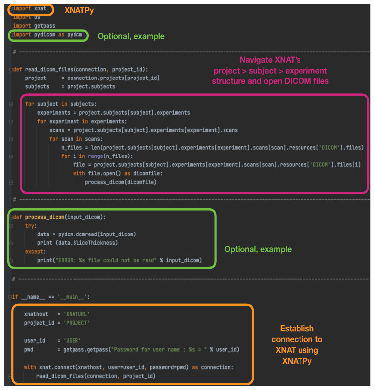
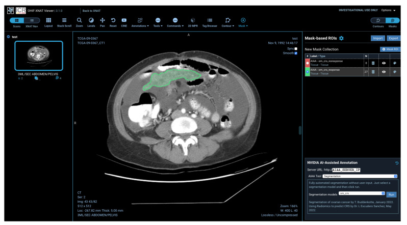
Artificial intelligence (AI) methods applied to healthcare problems have shown enormous potential to alleviate the burden of health services worldwide and to improve the accuracy and reproducibility of predictions. In particular, developments in computer vision are creating a paradigm shift in the analysis of radiological images, where AI tools are already capable of automatically detecting and precisely delineating tumours. However, such tools are generally developed in technical departments that continue to be siloed from where the real benefit would be achieved with their usage. Significant effort still needs to be made to make these advancements available, first in academic clinical research and ultimately in the clinical setting. In this paper, we demonstrate a prototype pipeline based entirely on open-source software and free of cost to bridge this gap, simplifying the integration of tools and models developed within the AI community into the clinical research setting, ensuring an accessible platform with visualisation applications that allow end-users such as radiologists to view and interact with the outcome of these AI tools.

摘要

应用于医疗保健问题的人工智能(AI)方法已显示出巨大潜力,可减轻全球卫生服务负担,并提高预测的准确性和可重复性。特别是,计算机视觉的发展正在给放射图像分析带来范式转变,AI工具已经能够自动检测并精确勾勒肿瘤。然而,此类工具通常是在技术部门开发的,这些部门与工具实际使用能产生真正效益的地方仍然相互隔离。仍需付出巨大努力,首先在学术临床研究中,最终在临床环境中,使这些进展得以应用。在本文中,我们展示了一个完全基于开源软件且免费的原型流程,以弥合这一差距,简化将AI社区内开发的工具和模型集成到临床研究环境中的过程,确保有一个可访问的平台,其可视化应用程序能让放射科医生等终端用户查看这些AI工具的结果并与之交互。

https://cdn.ncbi.nlm.nih.gov/pmc/blobs/8a76/10486639/752352369792/diagnostics-13-02813-g013.jpg
https://cdn.ncbi.nlm.nih.gov/pmc/blobs/8a76/10486639/4619d3c733fd/diagnostics-13-02813-g001.jpg
https://cdn.ncbi.nlm.nih.gov/pmc/blobs/8a76/10486639/7ed7ee3aedc0/diagnostics-13-02813-g002.jpg
https://cdn.ncbi.nlm.nih.gov/pmc/blobs/8a76/10486639/616031e2ca7c/diagnostics-13-02813-g003.jpg
https://cdn.ncbi.nlm.nih.gov/pmc/blobs/8a76/10486639/2619909dac34/diagnostics-13-02813-g004.jpg
https://cdn.ncbi.nlm.nih.gov/pmc/blobs/8a76/10486639/9a228940f3d4/diagnostics-13-02813-g005.jpg
https://cdn.ncbi.nlm.nih.gov/pmc/blobs/8a76/10486639/33ec038b0a26/diagnostics-13-02813-g006.jpg
https://cdn.ncbi.nlm.nih.gov/pmc/blobs/8a76/10486639/2a8239c75cc3/diagnostics-13-02813-g007.jpg
https://cdn.ncbi.nlm.nih.gov/pmc/blobs/8a76/10486639/5662e8f4838f/diagnostics-13-02813-g008.jpg
https://cdn.ncbi.nlm.nih.gov/pmc/blobs/8a76/10486639/4c1b23d3ff6c/diagnostics-13-02813-g009.jpg
https://cdn.ncbi.nlm.nih.gov/pmc/blobs/8a76/10486639/1bd57ef0c9e7/diagnostics-13-02813-g010.jpg
https://cdn.ncbi.nlm.nih.gov/pmc/blobs/8a76/10486639/dc8b7a0785f2/diagnostics-13-02813-g011.jpg
https://cdn.ncbi.nlm.nih.gov/pmc/blobs/8a76/10486639/f017f5c95b43/diagnostics-13-02813-g012.jpg
https://cdn.ncbi.nlm.nih.gov/pmc/blobs/8a76/10486639/752352369792/diagnostics-13-02813-g013.jpg

相似文献

1
Integrating Artificial Intelligence Tools in the Clinical Research Setting: The Ovarian Cancer Use Case.在临床研究环境中整合人工智能工具:卵巢癌应用案例
Diagnostics (Basel). 2023 Aug 30;13(17):2813. doi: 10.3390/diagnostics13172813.
2
Browser-based Data Annotation, Active Learning, and Real-Time Distribution of Artificial Intelligence Models: From Tumor Tissue Microarrays to COVID-19 Radiology.基于浏览器的数据标注、主动学习与人工智能模型的实时分发:从肿瘤组织微阵列到COVID-19放射学
J Pathol Inform. 2021 Sep 27;12:38. doi: 10.4103/jpi.jpi_100_20. eCollection 2021.
3
Artificial intelligence in radiography: Where are we now and what does the future hold?人工智能在放射学中的应用:我们现在在哪里,未来会怎样?
Radiography (Lond). 2021 Oct;27 Suppl 1:S58-S62. doi: 10.1016/j.radi.2021.07.015. Epub 2021 Aug 8.
4
Leveraging artificial intelligence and software engineering methods in epidemiology for the co-creation of decision-support tools based on mechanistic models.利用人工智能和软件工程方法在流行病学中共同创建基于机械模型的决策支持工具。
Prev Vet Med. 2024 Jul;228:106233. doi: 10.1016/j.prevetmed.2024.106233. Epub 2024 May 25.
5
The future of Cochrane Neonatal.考克兰新生儿协作网的未来。
Early Hum Dev. 2020 Nov;150:105191. doi: 10.1016/j.earlhumdev.2020.105191. Epub 2020 Sep 12.
6
How to prepare for a bright future of radiology in Europe.如何为欧洲放射学的光明未来做好准备。
Insights Imaging. 2023 Oct 10;14(1):168. doi: 10.1186/s13244-023-01525-3.
7
How Artificial Intelligence Is Shaping Medical Imaging Technology: A Survey of Innovations and Applications.人工智能如何塑造医学成像技术:创新与应用综述
Bioengineering (Basel). 2023 Dec 18;10(12):1435. doi: 10.3390/bioengineering10121435.
8
Emerging Trends in AI and Radiomics for Bladder, Kidney, and Prostate Cancer: A Critical Review.人工智能与放射组学在膀胱癌、肾癌和前列腺癌中的新趋势:批判性综述
Cancers (Basel). 2024 Feb 16;16(4):810. doi: 10.3390/cancers16040810.
9
Overview of Artificial Intelligence in Breast Cancer Medical Imaging.乳腺癌医学成像中的人工智能概述
J Clin Med. 2023 Jan 4;12(2):419. doi: 10.3390/jcm12020419.
10
A novel reporting workflow for automated integration of artificial intelligence results into structured radiology reports.一种将人工智能结果自动整合到结构化放射学报告中的新型报告工作流程。
Insights Imaging. 2024 Mar 19;15(1):80. doi: 10.1186/s13244-024-01660-5.

引用本文的文献

1
Future theranostic strategies: emerging ovarian cancer biomarkers to bridge the gap between diagnosis and treatment.未来的诊疗策略:新兴的卵巢癌生物标志物以弥合诊断与治疗之间的差距。
Front Drug Deliv. 2024 Feb 1;4:1339936. doi: 10.3389/fddev.2024.1339936. eCollection 2024.

本文引用的文献

1
Deep learning-based segmentation of multisite disease in ovarian cancer.基于深度学习的卵巢癌多部位病变分割。
Eur Radiol Exp. 2023 Dec 7;7(1):77. doi: 10.1186/s41747-023-00388-z.
2
Calibrating ensembles for scalable uncertainty quantification in deep learning-based medical image segmentation.基于深度学习的医学图像分割中可扩展不确定性量化的集成校准。
Comput Biol Med. 2023 Sep;163:107096. doi: 10.1016/j.compbiomed.2023.107096. Epub 2023 Jun 1.
3
Automated segmentation of colorectal liver metastasis and liver ablation on contrast-enhanced CT images.
基于对比增强CT图像的结直肠癌肝转移灶自动分割及肝脏消融
Front Oncol. 2022 Aug 11;12:886517. doi: 10.3389/fonc.2022.886517. eCollection 2022.
4
Clinically Interpretable Radiomics-Based Prediction of Histopathologic Response to Neoadjuvant Chemotherapy in High-Grade Serous Ovarian Carcinoma.基于影像组学的高级别浆液性卵巢癌新辅助化疗组织病理学反应的临床可解释预测
Front Oncol. 2022 Jun 16;12:868265. doi: 10.3389/fonc.2022.868265. eCollection 2022.
5
Machine learning for medical imaging: methodological failures and recommendations for the future.医学成像中的机器学习:方法学上的失败与未来建议。
NPJ Digit Med. 2022 Apr 12;5(1):48. doi: 10.1038/s41746-022-00592-y.
6
The difficulty of computing stable and accurate neural networks: On the barriers of deep learning and Smale's 18th problem.计算稳定且准确的神经网络的困难:深度学习与斯梅尔第 18 问题的障碍。
Proc Natl Acad Sci U S A. 2022 Mar 22;119(12):e2107151119. doi: 10.1073/pnas.2107151119. Epub 2022 Mar 16.
7
Integrating the OHIF Viewer into XNAT: Achievements, Challenges and Prospects for Quantitative Imaging Studies.将 OHIF 查看器集成到 XNAT 中:实现定量成像研究的成就、挑战和前景。
Tomography. 2022 Feb 11;8(1):497-512. doi: 10.3390/tomography8010040.
8
Automatic Segmentation of Kidneys and Kidney Tumors: The KiTS19 International Challenge.肾脏及肾肿瘤的自动分割:KiTS19国际挑战赛
Front Digit Health. 2022 Jan 4;3:797607. doi: 10.3389/fdgth.2021.797607. eCollection 2021.
9
A systematic review and quality of reporting checklist for repeatability and reproducibility of radiomic features.一项关于放射组学特征可重复性和再现性的系统评价及报告质量清单
Phys Imaging Radiat Oncol. 2021 Nov 9;20:69-75. doi: 10.1016/j.phro.2021.10.007. eCollection 2021 Oct.
10
Deep Learning for Brain Tumor Segmentation: A Survey of State-of-the-Art.用于脑肿瘤分割的深度学习:最新技术综述
J Imaging. 2021 Jan 29;7(2):19. doi: 10.3390/jimaging7020019.