Anderson Brian M, Rigaud Bastien, Lin Yuan-Mao, Jones A Kyle, Kang HynSeon Christine, Odisio Bruno C, Brock Kristy K
Department of Imaging Physics, The University of Texas MD Anderson Cancer Center, Houston, TX, United States.
UTHealth Graduate School of Biomedical Sciences, The University of Texas MD Anderson Cancer Center, Houston, TX, United States.
Front Oncol. 2022 Aug 11;12:886517. doi: 10.3389/fonc.2022.886517. eCollection 2022.
Colorectal cancer (CRC), the third most common cancer in the USA, is a leading cause of cancer-related death worldwide. Up to 60% of patients develop liver metastasis (CRLM). Treatments like radiation and ablation therapies require disease segmentation for planning and therapy delivery. For ablation, ablation-zone segmentation is required to evaluate disease coverage. We hypothesize that fully convolutional (FC) neural networks, trained using novel methods, will provide rapid and accurate identification and segmentation of CRLM and ablation zones.
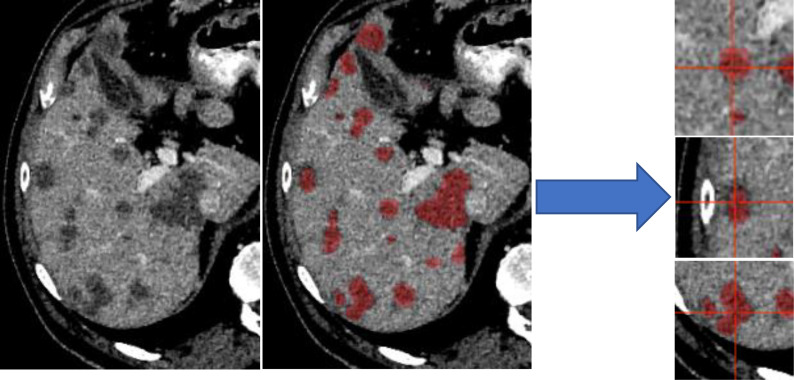
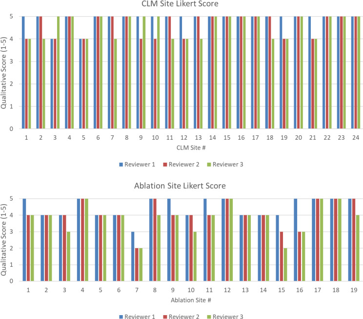
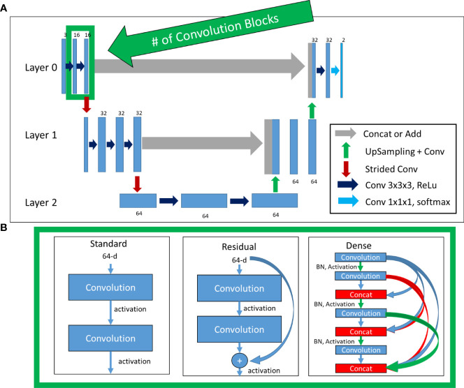
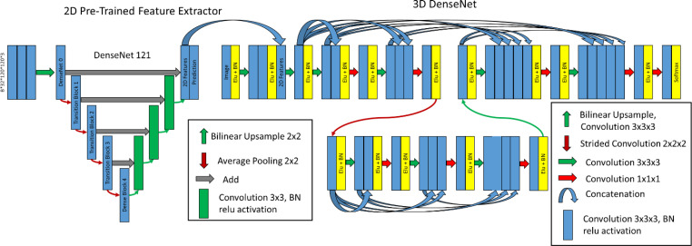
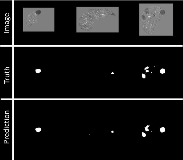
Four FC model styles were investigated: Standard 3D-UNet, Residual 3D-UNet, Dense 3D-UNet, and Hybrid-WNet. Models were trained on 92 patients from the liver tumor segmentation (LiTS) challenge. For the evaluation, we acquired 15 patients from the 3D-IRCADb database, 18 patients from our institution (CRLM = 24, ablation-zone = 19), and those submitted to the LiTS challenge ( = 70). Qualitative evaluations of our institutional data were performed by two board-certified radiologists (interventional and diagnostic) and a radiology-trained physician fellow, using a Likert scale of 1-5.
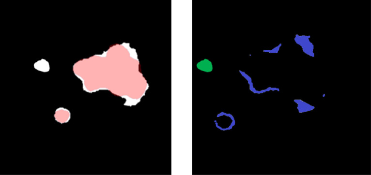
The most accurate model was the Hybrid-WNet. On a patient-by-patient basis in the 3D-IRCADb dataset, the median (min-max) Dice similarity coefficient (DSC) was 0.73 (0.41-0.88), the median surface distance was 1.75 mm (0.57-7.63 mm), and the number of false positives was 1 (0-4). In the LiTS challenge ( = 70), the global DSC was 0.810. The model sensitivity was 98% (47/48) for sites ≥15 mm in diameter. Qualitatively, 100% (24/24; minority vote) of the CRLM and 84% (16/19; majority vote) of the ablation zones had Likert scores ≥4.
The Hybrid-WNet model provided fast (<30 s) and accurate segmentations of CRLM and ablation zones on contrast-enhanced CT scans, with positive physician reviews.
结直肠癌(CRC)是美国第三大常见癌症,也是全球癌症相关死亡的主要原因。高达60%的患者会发生肝转移(CRLM)。放射治疗和消融治疗等需要对疾病进行分割以用于治疗计划和治疗实施。对于消融治疗,需要对消融区域进行分割以评估疾病覆盖范围。我们假设,使用新方法训练的全卷积(FC)神经网络将能快速、准确地识别和分割CRLM及消融区域。
研究了四种FC模型样式:标准3D - UNet、残差3D - UNet、密集3D - UNet和混合W - Net。模型在来自肝脏肿瘤分割(LiTS)挑战赛的92例患者数据上进行训练。为进行评估,我们从3D - IRCADb数据库获取了15例患者,从我们机构获取了18例患者(CRLM = 24例,消融区域 = 19例),以及提交给LiTS挑战赛的患者( = 70例)。由两位获得委员会认证的放射科医生(介入和诊断)以及一名经过放射学培训的医师对我们机构的数据进行定性评估,使用1 - 5的李克特量表。
最准确的模型是混合W - Net。在3D - IRCADb数据集中逐患者来看,中位(最小 - 最大)骰子相似系数(DSC)为0.73(0.41 - 0.88),中位表面距离为1.75毫米(0.57 - 7.63毫米),假阳性数量为1(0 - 4)。在LiTS挑战赛( = 70)中,全局DSC为0.810。对于直径≥15毫米的部位,模型敏感性为98%(47/48)。定性来看,100%(24/24;少数服从多数投票)的CRLM和84%(16/19;多数投票)的消融区域李克特评分≥4。
混合W - Net模型在增强CT扫描上能快速(<30秒)且准确地分割CRLM和消融区域,医师评价良好。