Suppr超能文献

经阴道三维超声在 11+3 至 13+6 孕周时对前脑结构、神经节隆起、基底节和丘脑的发育研究及其正常数据。

Development of the prosencephalic structures, ganglionic eminence, basal ganglia and thalamus at 11 + 3 to 13 + 6 gestational weeks on 3D transvaginal ultrasound including normative data.

机构信息

Department of Gynecology, Obstetrics and Gynecological Endocrinology, Johannes Kepler University Linz, Kepler University Hospital, Altenberger Strasse 69, 4040 Linz and Krankenhausstraße 26-30, 4020, Linz, Austria.

Department of Pediatrics and Adolescent Medicine, Johannes Kepler University Linz, Kepler University Hospital, Altenberger Strasse 69, 4040 Linz and Krankenhausstrasse 26-30, 4020, Linz, Austria.

出版信息

Brain Struct Funct. 2023 Dec;228(9):2089-2101. doi: 10.1007/s00429-023-02679-y. Epub 2023 Sep 15.

Abstract

OBJECTIVES

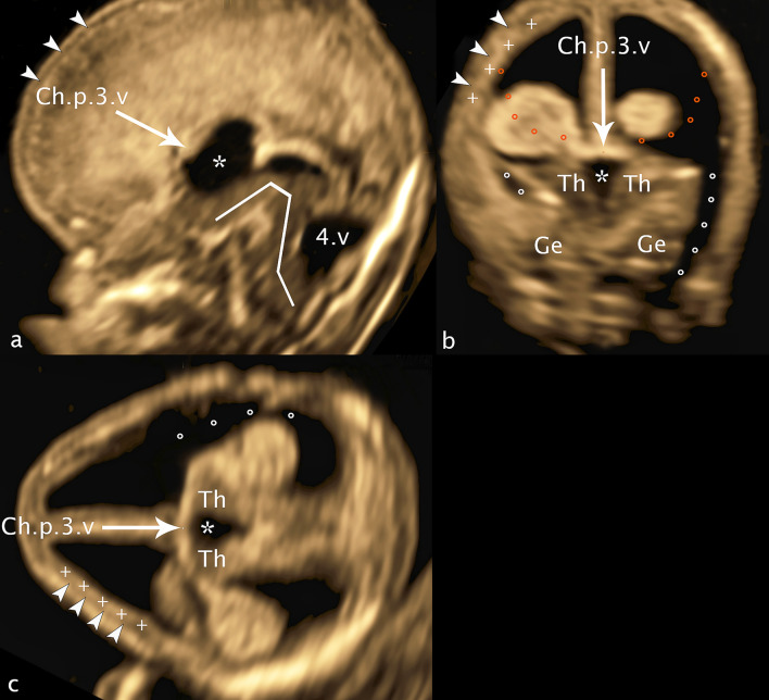
To show the development of ganglionic eminence, basal ganglia and thalamus/hypothalamus in week 11 + 3 to 13 + 6 by transvaginal 3D ultrasound.

METHODS

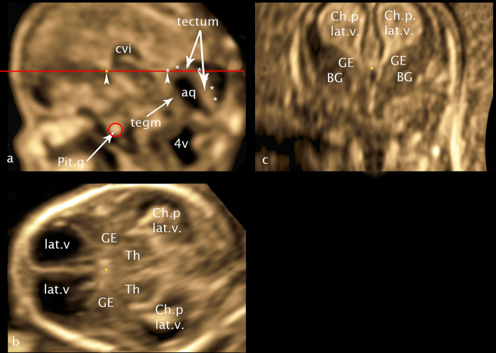
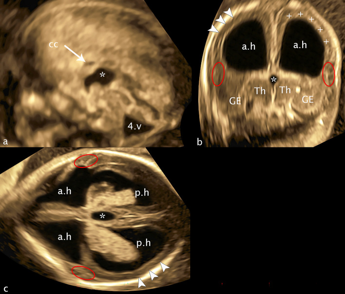
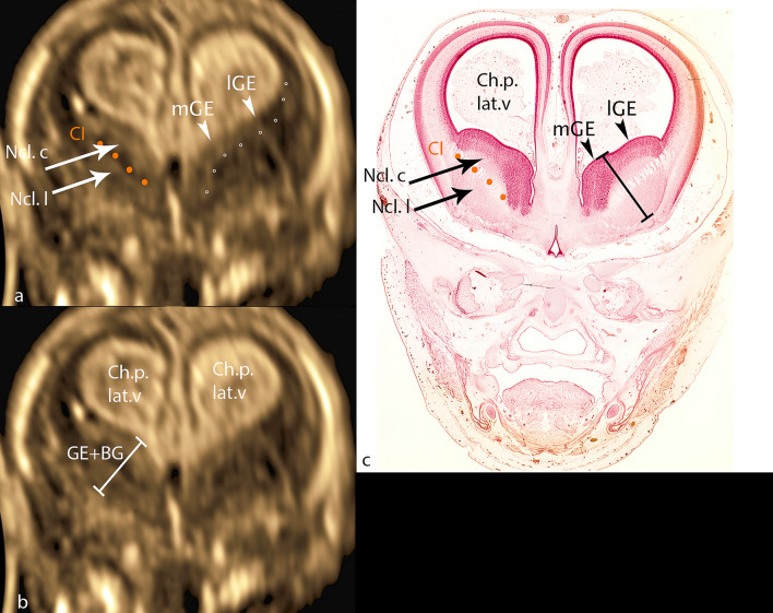
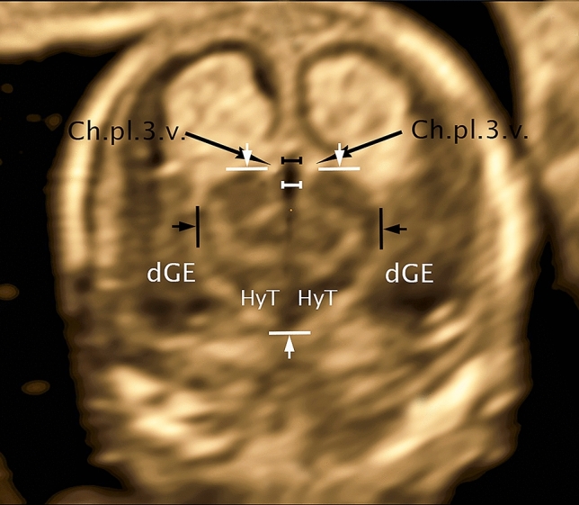
To visualize the prosencephalic structures surrounding the 3rd ventricle, 285 three-dimensional ultrasound volume blocks from 402 fetuses examined were selected in a prospective transvaginal 3D study to compare ultrasound images of ganglionic eminence, basal ganglia, thalamus/hypothalamus with embryological sections. In addition, measurements of the described structures were made in 104 fetuses to quantify the embryological development.

RESULTS

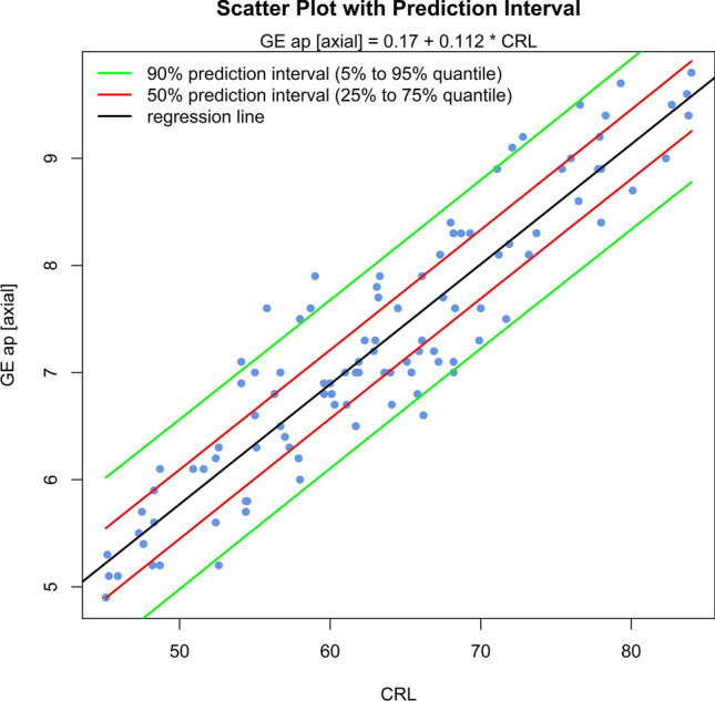
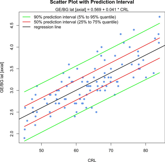
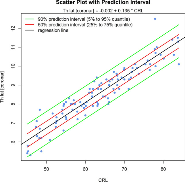
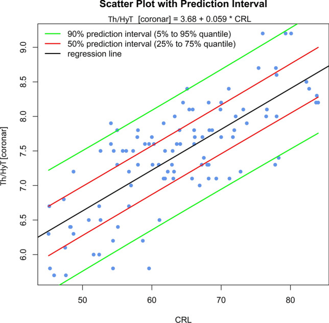
The sonomorphologic characteristics of ganglionic eminence, basal ganglia and thalamus/hypothalamus are described in 71% of the fetuses examined. Measurements of the structures in 57% of the fetuses, show the following results: axGE ap = 0.17 + 0.112CRL; axGE/I = 0.888 + 0.048CRL; axGE/BG = 0.569 + 0.041CRL; coGE/BG = 0.381 + 0.048CRL; coTh lat = - 0.002 + 0.135CRL; coTh/HyT = 3.68 + 0.059CRL; co3.V lat = 0.54 + 0.008*CRL.

CONCLUSION

Transvaginal 3D neurosonography allows visualization and measurement of normal structures in the fetal prosencephalon at 11 + 3 to 13 + 6 weeks of gestation (GW) including details of ganglionic eminence (GE), basal ganglia (BG), and thalamus/hypothalamus (Th/HyT). Further scientific work is needed before using the results to decide on pathological changes in patients.

摘要

目的

通过经阴道 3D 超声显示 11+3 周至 13+6 周胎儿的神经节隆起、基底节和丘脑/下丘脑的发育情况。

方法

为了观察第三脑室周围的前脑结构,在一项前瞻性经阴道 3D 研究中,从 402 例检查的胎儿中选择了 285 个三维超声容积块,将超声图像与胚胎学切片进行比较,以比较神经节隆起、基底节、丘脑/下丘脑的超声图像。此外,在 104 例胎儿中对描述的结构进行了测量,以量化胚胎发育情况。

结果

在 71%的检查胎儿中描述了神经节隆起、基底节和丘脑/下丘脑的超声形态特征。在 57%的胎儿中对结构进行了测量,结果如下:axGE ap=0.17+0.112CRL;axGE/I=0.888+0.048CRL;axGE/BG=0.569+0.041CRL;coGE/BG=0.381+0.048CRL;coTh lat=-0.002+0.135CRL;coTh/HyT=3.68+0.059CRL;co3.V lat=0.54+0.008*CRL。

结论

经阴道 3D 神经超声可在 11+3 周至 13+6 周妊娠(GW)时显示和测量胎儿前脑的正常结构,包括神经节隆起(GE)、基底节(BG)和丘脑/下丘脑(Th/HyT)的细节。在将结果用于决定患者的病理性变化之前,需要进一步的科学研究。

https://cdn.ncbi.nlm.nih.gov/pmc/blobs/7434/10587255/2529e32bca52/429_2023_2679_Fig1_HTML.jpg

相似文献

2
Normal and abnormal appearance of fetal ganglionic eminence on second-trimester three-dimensional ultrasound.
Ultrasound Obstet Gynecol. 2023 Sep;62(3):398-404. doi: 10.1002/uog.26229. Epub 2023 Aug 8.
4
Ganglionic eminence within the early developing brain visualized by 3D transvaginal ultrasound.
Med Ultrason. 2015 Sep;17(3):289-94. doi: 10.11152/mu.2013.2066.173.rbb.
5
Reference Ranges for Transvaginal Examined Fossa Posterior Structures in Fetuses from 45 to 84 mm Crown-Rump Length.
Gynecol Obstet Invest. 2018;83(4):375-380. doi: 10.1159/000486334. Epub 2018 Jun 5.
7
Transvaginal three-dimensional sonographic assessment of the embryonic brain: a pilot study.
Clujul Med. 2015;88(2):152-8. doi: 10.15386/cjmed-437. Epub 2015 Apr 15.

引用本文的文献

1
Morphometry of the Human Ventral Germinal Matrix during Fetal Development.
Bull Exp Biol Med. 2025 Mar;178(5):675-680. doi: 10.1007/s10517-025-06396-w. Epub 2025 Apr 29.

本文引用的文献

1
Fetal Brain Damage in Human Fetuses with Congenital Cytomegalovirus Infection: Histological Features and Viral Tropism.
Cell Mol Neurobiol. 2023 Apr;43(3):1385-1399. doi: 10.1007/s10571-022-01258-9. Epub 2022 Aug 7.
2
Sonography of fetal holoprosencephaly: a guide to recognize the lesser varieties.
J Matern Fetal Neonatal Med. 2022 Dec;35(25):9717-9723. doi: 10.1080/14767058.2022.2050900. Epub 2022 Mar 10.
5
First-trimester fetal neurosonography: technique and diagnostic potential.
Ultrasound Obstet Gynecol. 2021 Feb;57(2):204-214. doi: 10.1002/uog.23149.
7
Ratio of fetal choroid plexus to head size: simple sonographic marker of open spina bifida at 11-13 weeks' gestation.
Ultrasound Obstet Gynecol. 2020 Jan;55(1):81-86. doi: 10.1002/uog.20856. Epub 2019 Dec 12.
8
Congenital Aqueductal Stenosis: Findings at Fetal MRI That Accurately Predict a Postnatal Diagnosis.
AJNR Am J Neuroradiol. 2018 May;39(5):942-948. doi: 10.3174/ajnr.A5590. Epub 2018 Mar 8.
9
Prenatal Diagnosis of Lissencephaly Type 2 using Three-dimensional Ultrasound and Fetal MRI: Case Report and Review of the Literature.
Rev Bras Ginecol Obstet. 2016 Apr;38(4):201-6. doi: 10.1055/s-0036-1582126. Epub 2016 Apr 18.
10
Ganglionic eminence within the early developing brain visualized by 3D transvaginal ultrasound.
Med Ultrason. 2015 Sep;17(3):289-94. doi: 10.11152/mu.2013.2066.173.rbb.

文献AI研究员

20分钟写一篇综述,助力文献阅读效率提升50倍。

立即体验

用中文搜PubMed

大模型驱动的PubMed中文搜索引擎

马上搜索

文档翻译

学术文献翻译模型,支持多种主流文档格式。

立即体验