Cao Ye, Kalajzic Ivo, Matthews Brya G
Department of Molecular Medicine and Pathology, University of Auckland, Auckland, New Zealand.
Center for Regenerative Medicine and Skeletal Development, School of Dental Medicine, UConn Health, Farmington, CT, United States.
Front Physiol. 2023 Sep 4;14:1231352. doi: 10.3389/fphys.2023.1231352. eCollection 2023.
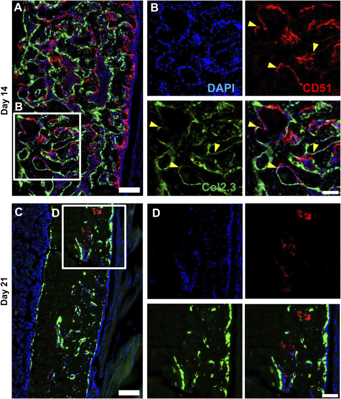
The periosteum is a critical source of skeletal stem and progenitor cells (SSPCs) that form callus tissue in response to injury. There is yet to be a consensus on how to identify SSPCs in the adult periosteum. The aim of this study was to understand how potential murine periosteal SSPC populations behave and in response to injury. We evaluated the differentiation potential of Sca1CD51 and Sca1CD51 cells following transplantation. , the Sca1CD51 population appears to be more primitive multipotent cells, but after transplantation, Sca1CD51 cells showed superior engraftment, expansion, and differentiation into chondrocytes and osteoblasts. Despite representing a clear population with flow cytometry, we identified very few Sca1CD51 cells histologically. Using a periosteal scratch injury model, we successfully mimicked the endochondral-like healing process seen in unstable fractures, including the expansion and osteochondral differentiation of αSMA cells following injury. CD51 cells were present in the cambium layer of resting periosteum and expanded following injury. Sca1CD51 cells were mainly localized in the outer periosteal layer. We found that injury increased colony-forming unit fibroblast (CFU-F) formation in the periosteum and led to rapid expansion of CD90 cells. Several other populations, including Sca1CD51 and CD34 cells, were expanded by day 7. Mice with enhanced fracture healing due to elevated Notch signaling mediated by NICD1 overexpression showed significant expansion of CD51 and CD34 cells in the early stages of healing, suggesting these populations contribute to more rapid healing. In conclusion, we demonstrate that periosteal injury leads to the expansion of various SSPC populations, but further studies are required to confirm their lineage hierarchy in the adult skeletal system. Our data indicate that CD51 skeletal progenitor cells are injury-responsive and show good engraftment and differentiation potential upon transplantation.
骨膜是骨骼干细胞和祖细胞(SSPCs)的关键来源,这些细胞在损伤后会形成骨痂组织。目前对于如何在成年骨膜中识别SSPCs尚未达成共识。本研究的目的是了解潜在的小鼠骨膜SSPC群体的行为以及对损伤的反应。我们评估了移植后Sca1CD51和Sca1CD51细胞的分化潜能。结果显示,Sca1CD51群体似乎是更原始的多能细胞,但移植后,Sca1CD51细胞表现出更好的植入、增殖以及向软骨细胞和成骨细胞分化的能力。尽管通过流式细胞术可明确区分该群体,但在组织学上我们仅发现极少数Sca1CD51细胞。利用骨膜刮擦损伤模型,我们成功模拟了不稳定骨折中所见的软骨内样愈合过程,包括损伤后αSMA细胞的增殖和骨软骨分化。CD51细胞存在于静止骨膜的生发层,并在损伤后增殖。Sca1CD51细胞主要定位于骨膜外层。我们发现损伤增加了骨膜中集落形成单位成纤维细胞(CFU-F)的形成,并导致CD90细胞迅速增殖。到第7天时,包括Sca1CD51和CD34细胞在内的其他几个群体也出现增殖。由于NICD1过表达介导的Notch信号增强而导致骨折愈合加快的小鼠,在愈合早期CD51和CD34细胞显著增殖,表明这些群体有助于更快愈合。总之,我们证明骨膜损伤会导致各种SSPC群体的增殖,但需要进一步研究来确认它们在成年骨骼系统中的谱系层次。我们的数据表明,CD51骨骼祖细胞对损伤有反应,移植后具有良好的植入和分化潜能。