Verrecchia-Ramos Emilie, Morel Olivier, Ginet Merwan, Retif Paul, Ben Mahmoud Sinan
Department of Medical Physics, Mercy Hospital, CHR Metz-Thionville, 1, Allée du Château, 57530, Ars-Laquenexy, France.
Department of Nuclear Medicine, Mercy Hospital, CHR Metz-Thionville, 1, Allée du Château, 57530, Ars-Laquenexy, France.
EJNMMI Phys. 2023 Sep 21;10(1):57. doi: 10.1186/s40658-023-00578-z.
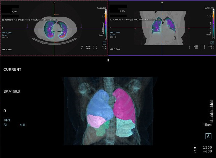
Lung lobar ventilation and perfusion (V/Q) quantification is generally obtained by generating planar scintigraphy images and then imposing three equally sized regions of interest on the data of each lung. This method is fast but not as accurate as SPECT/CT imaging, which provides three-dimensional data and therefore allows more precise lobar quantification. However, the manual delineation of each lobe is time-consuming, which makes SPECT/CT incompatible with the clinical workflow for V/Q estimation. An alternative may be to use artificial intelligence-based auto-segmentation tools such as AutoLung3D (Siemens Healthineers, Knoxville, USA), which automatically delineate the lung lobes on the CT data acquired with the SPECT data. The present study assessed the clinical validity of this approach relative to planar scintigraphy and manual quantification in SPECT/CT.
The Autolung3D software was tested on the retrospective SPECT/CT data of 43 patients who underwent V/Q scintigraphy with Tc-macroaggregated albumin and Tc-labeled aerosol. It was compared to planar scintigraphy and SPECT/CT using the manual quantification method in terms of relative lobar V/Q quantification values and interobserver variability.
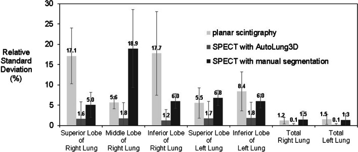
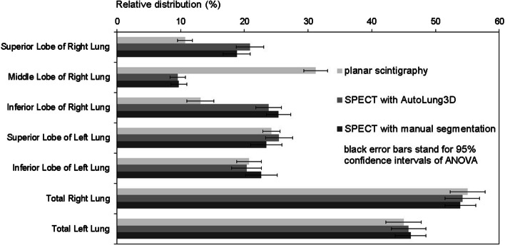
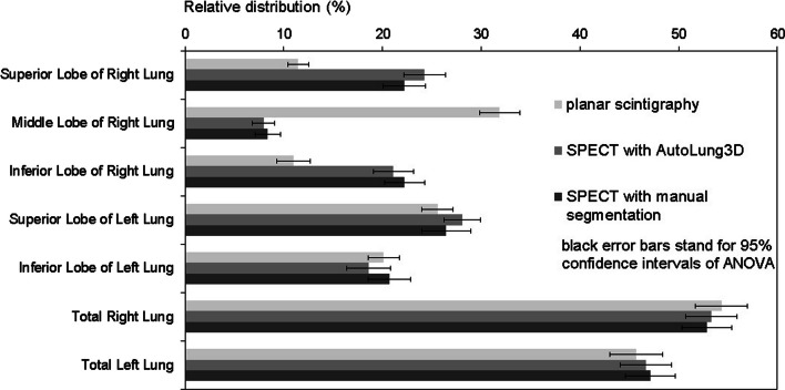
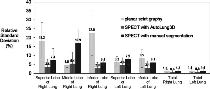
The three methods provided similar V/Q estimates for the left lung lobes and total lungs. However, compared to the manual SPECT/CT method, planar scintigraphy yielded significantly higher estimates for the middle right lobe and significantly lower estimates for the superior and inferior right lobes. The estimates of the manual and automated SPECT/CT methods were similar. However, the post-processing time in the automated method was approximately 5 min compared to 2 h for the manual method. Moreover, the automated method associated with a drastic reduction in interobserver variability: Its maximal relative standard deviation was only 5%, compared to 23% for planar scintigraphy and 19% for the manual SPECT/CT method.
This study validated the AutoLung3D software for general clinical use since it rapidly provides accurate lobar quantification in V/Q scans with markedly less interobserver variability than planar scintigraphy or the manual SPECT/CT method.
肺叶通气与灌注(V/Q)定量通常通过生成平面闪烁显像图像,然后在每个肺的数据上设置三个大小相等的感兴趣区来获得。这种方法速度快,但不如SPECT/CT成像准确,SPECT/CT成像可提供三维数据,因此能实现更精确的肺叶定量。然而,手动勾勒每个肺叶耗时,这使得SPECT/CT与V/Q评估的临床工作流程不兼容。一种替代方法可能是使用基于人工智能的自动分割工具,如AutoLung3D(西门子医疗,美国诺克斯维尔),它能在与SPECT数据一起采集的CT数据上自动勾勒肺叶。本研究评估了这种方法相对于平面闪烁显像和SPECT/CT中手动定量的临床有效性。
使用AutoLung3D软件对43例行V/Q闪烁显像(使用Tc-大颗粒聚合白蛋白和Tc标记气雾剂)的患者的回顾性SPECT/CT数据进行测试。在相对肺叶V/Q定量值和观察者间变异性方面,将其与平面闪烁显像和采用手动定量方法的SPECT/CT进行比较。
三种方法对左肺叶和全肺的V/Q估计相似。然而,与手动SPECT/CT方法相比,平面闪烁显像对右中叶的估计值显著更高,对右上叶和右下叶的估计值显著更低。手动和自动SPECT/CT方法的估计值相似。然而,自动方法的后处理时间约为5分钟,而手动方法为2小时。此外,自动方法使观察者间变异性大幅降低:其最大相对标准差仅为5%,相比之下,平面闪烁显像为23%,手动SPECT/CT方法为19%。
本研究验证了AutoLung3D软件可用于一般临床,因为它能在V/Q扫描中快速提供准确的肺叶定量,且观察者间变异性明显小于平面闪烁显像或手动SPECT/CT方法。