• 文献检索
  • 文档翻译
  • 深度研究
  • 学术资讯
  • Suppr Zotero 插件Zotero 插件
  • 邀请有礼
  • 套餐&价格
  • 历史记录
应用&插件
Suppr Zotero 插件Zotero 插件浏览器插件Mac 客户端Windows 客户端微信小程序
定价
高级版会员购买积分包购买API积分包
服务
文献检索文档翻译深度研究API 文档MCP 服务
关于我们
关于 Suppr公司介绍联系我们用户协议隐私条款
关注我们

Suppr 超能文献

核心技术专利:CN118964589B侵权必究
粤ICP备2023148730 号-1Suppr @ 2026

文献检索

告别复杂PubMed语法,用中文像聊天一样搜索,搜遍4000万医学文献。AI智能推荐,让科研检索更轻松。

立即免费搜索

文件翻译

保留排版,准确专业,支持PDF/Word/PPT等文件格式,支持 12+语言互译。

免费翻译文档

深度研究

AI帮你快速写综述,25分钟生成高质量综述,智能提取关键信息,辅助科研写作。

立即免费体验

使用深度卷积神经网络对超声图像中的乳腺病变进行分类:迁移学习与自动架构设计。

Classification of breast lesions in ultrasound images using deep convolutional neural networks: transfer learning versus automatic architecture design.

机构信息

School of Computing and Engineering, University of Derby, Derby, DE22 3AW, UK.

Department of Ultrasound, Shuguang Hospital affiliated to Shanghai University of Traditional Chinese Medicine, Shanghai, China.

出版信息

Med Biol Eng Comput. 2024 Jan;62(1):135-149. doi: 10.1007/s11517-023-02922-y. Epub 2023 Sep 22.

DOI:10.1007/s11517-023-02922-y
PMID:37735296
原文链接:https://pmc.ncbi.nlm.nih.gov/articles/PMC10758370/
Abstract

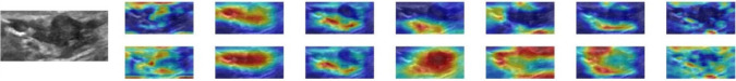
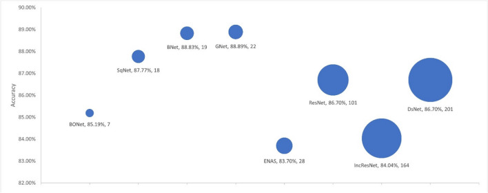
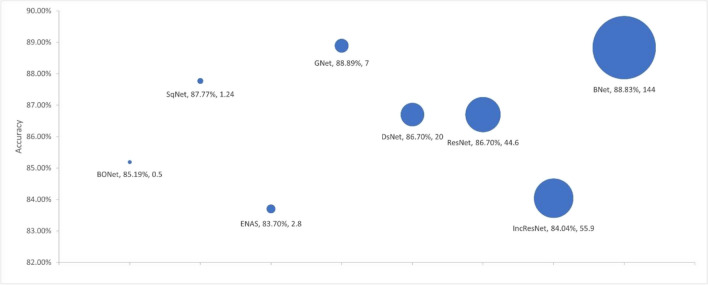
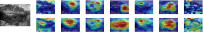
Deep convolutional neural networks (DCNNs) have demonstrated promising performance in classifying breast lesions in 2D ultrasound (US) images. Exiting approaches typically use pre-trained models based on architectures designed for natural images with transfer learning. Fewer attempts have been made to design customized architectures specifically for this purpose. This paper presents a comprehensive evaluation on transfer learning based solutions and automatically designed networks, analyzing the accuracy and robustness of different recognition models in three folds. First, we develop six different DCNN models (BNet, GNet, SqNet, DsNet, RsNet, IncReNet) based on transfer learning. Second, we adapt the Bayesian optimization method to optimize a CNN network (BONet) for classifying breast lesions. A retrospective dataset of 3034 US images collected from various hospitals is then used for evaluation. Extensive tests show that the BONet outperforms other models, exhibiting higher accuracy (83.33%), lower generalization gap (1.85%), shorter training time (66 min), and less model complexity (approximately 0.5 million weight parameters). We also compare the diagnostic performance of all models against that by three experienced radiologists. Finally, we explore the use of saliency maps to explain the classification decisions made by different models. Our investigation shows that saliency maps can assist in comprehending the classification decisions.

摘要

深度卷积神经网络(DCNN)在二维超声(US)图像中对乳腺病变的分类表现出了很有前景的性能。现有的方法通常使用基于自然图像架构的预训练模型进行迁移学习。针对这一目的,设计定制架构的尝试较少。本文对基于迁移学习的解决方案和自动设计的网络进行了全面评估,从三个方面分析了不同识别模型的准确性和鲁棒性。首先,我们基于迁移学习开发了六个不同的 DCNN 模型(BNet、GNet、SqNet、DsNet、RsNet、IncReNet)。其次,我们采用贝叶斯优化方法来优化用于分类乳腺病变的 CNN 网络(BONet)。然后,我们使用来自不同医院的 3034 个 US 图像的回顾性数据集进行评估。大量测试表明,BONet 优于其他模型,表现出更高的准确性(83.33%)、更低的泛化差距(1.85%)、更短的训练时间(66 分钟)和更少的模型复杂度(约 50 万个权重参数)。我们还比较了所有模型与三位经验丰富的放射科医生的诊断性能。最后,我们探索了使用显着性图来解释不同模型的分类决策。我们的研究表明,显着性图可以帮助理解分类决策。

https://cdn.ncbi.nlm.nih.gov/pmc/blobs/86db/10758370/c7b0c829fa1e/11517_2023_2922_Fig11_HTML.jpg
https://cdn.ncbi.nlm.nih.gov/pmc/blobs/86db/10758370/1beb4ed51b13/11517_2023_2922_Fig1_HTML.jpg
https://cdn.ncbi.nlm.nih.gov/pmc/blobs/86db/10758370/70be2903ce3a/11517_2023_2922_Fig2_HTML.jpg
https://cdn.ncbi.nlm.nih.gov/pmc/blobs/86db/10758370/0fef42f14b4e/11517_2023_2922_Fig3_HTML.jpg
https://cdn.ncbi.nlm.nih.gov/pmc/blobs/86db/10758370/fb0c4485ecc6/11517_2023_2922_Fig4_HTML.jpg
https://cdn.ncbi.nlm.nih.gov/pmc/blobs/86db/10758370/070192284e7a/11517_2023_2922_Fig5_HTML.jpg
https://cdn.ncbi.nlm.nih.gov/pmc/blobs/86db/10758370/4d6d29040655/11517_2023_2922_Fig6_HTML.jpg
https://cdn.ncbi.nlm.nih.gov/pmc/blobs/86db/10758370/08352e90a3e9/11517_2023_2922_Fig7_HTML.jpg
https://cdn.ncbi.nlm.nih.gov/pmc/blobs/86db/10758370/93777ed972bf/11517_2023_2922_Fig8_HTML.jpg
https://cdn.ncbi.nlm.nih.gov/pmc/blobs/86db/10758370/187667957007/11517_2023_2922_Fig9_HTML.jpg
https://cdn.ncbi.nlm.nih.gov/pmc/blobs/86db/10758370/16e7b39801c7/11517_2023_2922_Fig10_HTML.jpg
https://cdn.ncbi.nlm.nih.gov/pmc/blobs/86db/10758370/c7b0c829fa1e/11517_2023_2922_Fig11_HTML.jpg

相似文献

1
Classification of breast lesions in ultrasound images using deep convolutional neural networks: transfer learning versus automatic architecture design.使用深度卷积神经网络对超声图像中的乳腺病变进行分类:迁移学习与自动架构设计。
Med Biol Eng Comput. 2024 Jan;62(1):135-149. doi: 10.1007/s11517-023-02922-y. Epub 2023 Sep 22.
2
A generic deep learning framework to classify thyroid and breast lesions in ultrasound images.一种用于对超声图像中的甲状腺和乳腺病变进行分类的通用深度学习框架。
Ultrasonics. 2021 Feb;110:106300. doi: 10.1016/j.ultras.2020.106300. Epub 2020 Nov 12.
3
Explainable DCNN Decision Framework for Breast Lesion Classification from Ultrasound Images Based on Cancer Characteristics.基于癌症特征的超声图像乳腺病变分类可解释深度卷积神经网络决策框架
Bioengineering (Basel). 2024 May 2;11(5):453. doi: 10.3390/bioengineering11050453.
4
ENAS-B: Combining ENAS With Bayesian Optimization for Automatic Design of Optimal CNN Architectures for Breast Lesion Classification From Ultrasound Images.ENAS-B:将ENAS与贝叶斯优化相结合,用于从超声图像自动设计用于乳腺病变分类的最优卷积神经网络架构。
Ultrason Imaging. 2024 Jan;46(1):17-28. doi: 10.1177/01617346231208709. Epub 2023 Nov 20.
5
Automatic classification of ultrasound breast lesions using a deep convolutional neural network mimicking human decision-making.使用模仿人类决策的深度卷积神经网络对超声乳腺病变进行自动分类。
Eur Radiol. 2019 Oct;29(10):5458-5468. doi: 10.1007/s00330-019-06118-7. Epub 2019 Mar 29.
6
AutoTune: Automatically Tuning Convolutional Neural Networks for Improved Transfer Learning.AutoTune:自动调整卷积神经网络以提高迁移学习性能。
Neural Netw. 2021 Jan;133:112-122. doi: 10.1016/j.neunet.2020.10.009. Epub 2020 Oct 27.
7
Fus2Net: a novel Convolutional Neural Network for classification of benign and malignant breast tumor in ultrasound images.Fus2Net:一种用于超声图像中良性和恶性乳腺肿瘤分类的新型卷积神经网络。
Biomed Eng Online. 2021 Nov 18;20(1):112. doi: 10.1186/s12938-021-00950-z.
8
Comparison of Deep-Learning and Conventional Machine-Learning Methods for the Automatic Recognition of the Hepatocellular Carcinoma Areas from Ultrasound Images.深度学习与传统机器学习方法在自动识别超声图像肝癌区域的比较。
Sensors (Basel). 2020 May 29;20(11):3085. doi: 10.3390/s20113085.
9
Digital breast tomosynthesis versus digital mammography: integration of image modalities enhances deep learning-based breast mass classification.数字乳腺断层合成与数字乳腺钼靶摄影:图像模式的整合增强了基于深度学习的乳腺肿块分类。
Eur Radiol. 2020 Feb;30(2):778-788. doi: 10.1007/s00330-019-06457-5. Epub 2019 Nov 5.
10
A comparative study of pre-trained convolutional neural networks for semantic segmentation of breast tumors in ultrasound.用于超声乳腺肿瘤语义分割的预训练卷积神经网络的比较研究
Comput Biol Med. 2020 Nov;126:104036. doi: 10.1016/j.compbiomed.2020.104036. Epub 2020 Oct 8.

引用本文的文献

1
A Dirichlet Distribution-Based Complex Ensemble Approach for Breast Cancer Classification from Ultrasound Images with Transfer Learning and Multiphase Spaced Repetition Method.一种基于狄利克雷分布的复杂集成方法,用于通过迁移学习和多相间隔重复方法从超声图像中进行乳腺癌分类。
J Imaging Inform Med. 2025 Apr 29. doi: 10.1007/s10278-025-01515-5.
2
A multi-patch-based deep learning model with VGG19 for breast cancer classifications in the pathology images.一种基于多补丁的深度学习模型,采用VGG19用于病理图像中的乳腺癌分类。
Digit Health. 2025 Jan 17;11:20552076241313161. doi: 10.1177/20552076241313161. eCollection 2025 Jan-Dec.
3

本文引用的文献

1
Biphasic majority voting-based comparative COVID-19 diagnosis using chest X-ray images.基于双相多数投票的使用胸部X光图像的COVID-19比较诊断
Expert Syst Appl. 2023 Apr 15;216:119430. doi: 10.1016/j.eswa.2022.119430. Epub 2022 Dec 21.
2
Fus2Net: a novel Convolutional Neural Network for classification of benign and malignant breast tumor in ultrasound images.Fus2Net:一种用于超声图像中良性和恶性乳腺肿瘤分类的新型卷积神经网络。
Biomed Eng Online. 2021 Nov 18;20(1):112. doi: 10.1186/s12938-021-00950-z.
3
A generic deep learning framework to classify thyroid and breast lesions in ultrasound images.
Exploring the use of deep learning models for accurate tracking of 3D zebrafish trajectories.
探索使用深度学习模型精确跟踪三维斑马鱼轨迹。
Front Bioeng Biotechnol. 2024 Sep 25;12:1461264. doi: 10.3389/fbioe.2024.1461264. eCollection 2024.
4
Explainable DCNN Decision Framework for Breast Lesion Classification from Ultrasound Images Based on Cancer Characteristics.基于癌症特征的超声图像乳腺病变分类可解释深度卷积神经网络决策框架
Bioengineering (Basel). 2024 May 2;11(5):453. doi: 10.3390/bioengineering11050453.
5
A Review of Artificial Intelligence in Breast Imaging.人工智能在乳腺成像中的应用综述。
Tomography. 2024 May 9;10(5):705-726. doi: 10.3390/tomography10050055.
一种用于对超声图像中的甲状腺和乳腺病变进行分类的通用深度学习框架。
Ultrasonics. 2021 Feb;110:106300. doi: 10.1016/j.ultras.2020.106300. Epub 2020 Nov 12.
4
Colorectal cancer statistics, 2020.2020 年结直肠癌统计数据。
CA Cancer J Clin. 2020 May;70(3):145-164. doi: 10.3322/caac.21601. Epub 2020 Mar 5.
5
Breast Cancer Classification in Automated Breast Ultrasound Using Multiview Convolutional Neural Network with Transfer Learning.基于多视图卷积神经网络和迁移学习的自动乳腺超声乳腺癌分类。
Ultrasound Med Biol. 2020 May;46(5):1119-1132. doi: 10.1016/j.ultrasmedbio.2020.01.001. Epub 2020 Feb 12.
6
Dataset of breast ultrasound images.乳腺超声图像数据集。
Data Brief. 2019 Nov 21;28:104863. doi: 10.1016/j.dib.2019.104863. eCollection 2020 Feb.
7
Breast mass classification in sonography with transfer learning using a deep convolutional neural network and color conversion.超声中基于迁移学习的深度卷积神经网络和颜色转换的乳腺肿块分类。
Med Phys. 2019 Feb;46(2):746-755. doi: 10.1002/mp.13361. Epub 2019 Jan 16.
8
Comparison of Transferred Deep Neural Networks in Ultrasonic Breast Masses Discrimination.转移深度神经网络在超声乳腺肿块鉴别中的比较。
Biomed Res Int. 2018 Jun 21;2018:4605191. doi: 10.1155/2018/4605191. eCollection 2018.
9
Breast imaging reporting and data system (BI-RADS).乳腺影像报告和数据系统(BI-RADS)。
Radiol Clin North Am. 2002 May;40(3):409-30, v. doi: 10.1016/s0033-8389(01)00017-3.