Pediatric Surgery Department, Hospital Universitario de Navarra, Calle Irunlarrea 3, 31008, Pamplona, Navarra, Spain.
Preventive Medicine and Public Health Department, School of Medicine, University of Navarra, Pamplona, Navarra, Spain.
Pediatr Surg Int. 2023 Sep 22;39(1):274. doi: 10.1007/s00383-023-05558-z.
Scientific literature regarding the characterization of lymphocyte subpopulations of the cecal appendix is sparse, with few precedents limited to immunohistochemical techniques.
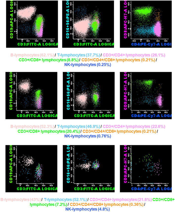
We conducted a prospective pilot study to characterize lymphocyte subpopulations of the cecal appendix in children. Participants were divided into three groups: (1) patients without histological acute appendiceal inflammation, (2) patients with histological uncomplicated acute appendicitis, and (3) patients with histological complicated acute appendicitis (gangrenous, perforated). A fresh sample of the base of the appendix was taken from all patients and a flow cytometric study was performed. Quantitative variables were compared using Kruskal-Wallis test and Mann-Whitney U test.
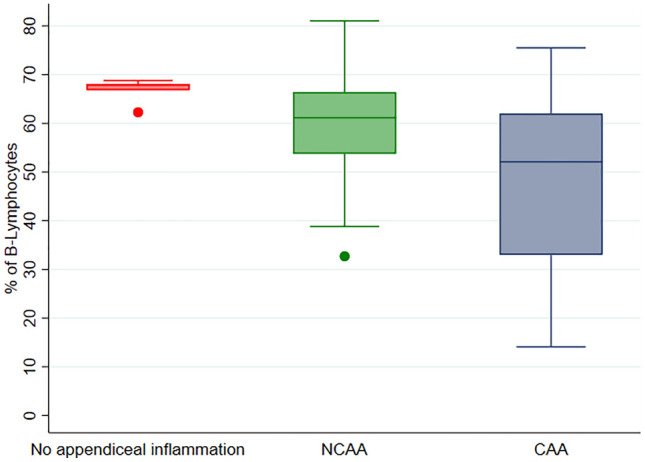
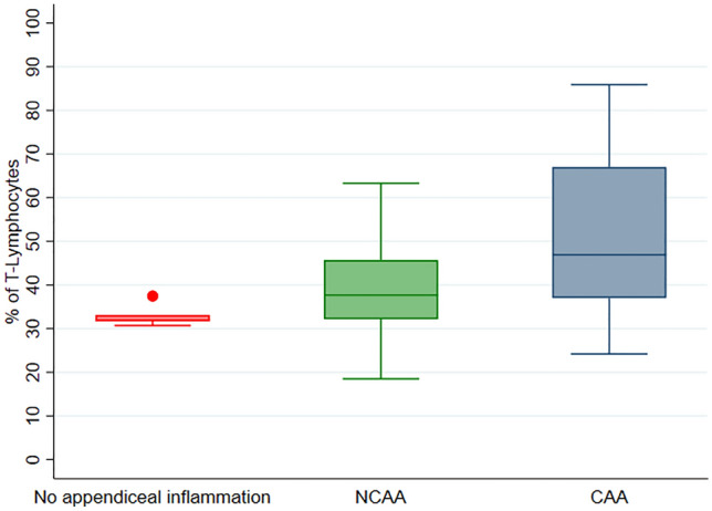
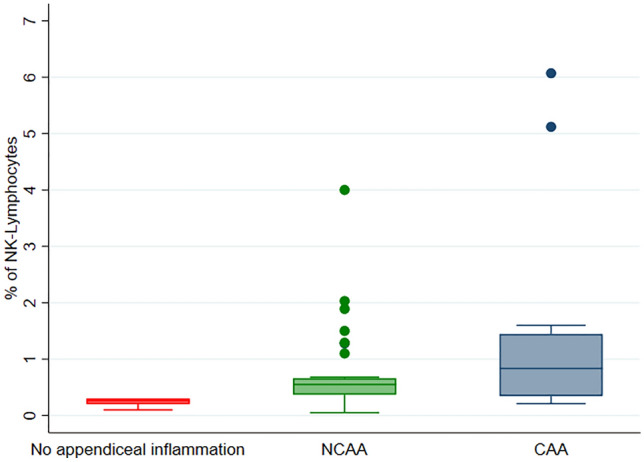
This study included 57 patients divided into Group 1 (n = 5), Group 2 (n = 37), and Group 3 (n = 15). Median values (IQR) of the percentage of B-lymphocytes were 67.8 [66.8-68.1] in group 1, 61.15 [53.74-66.4] in group 2, and 52.1 [33-62.02] in group 3 (p = 0.02). Median values (IQR) of the percentage of NK-lymphocytes were 0.26 [0.2-0.3] in group 1, 0.55 [0.37-0.66] in group 2, and 0.84 [0.35-1.45] in group 3 (p = 0.008). Median values (IQR) of the percentage of T-lymphocytes were 31.9 [31.7-33.1] in group 1, 37.68 [32.15-45.69] in group 2, and 46.9 [37.03-67] in group 3 (p = 0.02). Pair comparisons of groups 2 and 3 also showed significant differences in the percentage of B lymphocytes (p = 0.03) and NK-lymphocytes (p = 0.02).
Significant differences in lymphocyte subpopulations were identified according to the histologic grade of the cecal appendix. More specifically, a lower percentage of B-lymphocytes and a higher percentage of T- and NK-lymphocytes were observed in cases of acute appendicitis. These findings must be confirmed and their etiopathogenic, diagnostic, and prognostic implications elucidated in future studies with larger sample sizes.
关于盲肠阑尾淋巴细胞亚群特征的科学文献很少,只有少数先例仅限于免疫组织化学技术。
我们进行了一项前瞻性的初步研究,以描述儿童盲肠阑尾的淋巴细胞亚群。参与者分为三组:(1)无组织学急性阑尾炎的患者,(2)有组织学单纯性急性阑尾炎的患者,和(3)有组织学复杂性急性阑尾炎(坏疽性、穿孔性)的患者。所有患者均取自阑尾基底的新鲜样本,并进行流式细胞术研究。使用 Kruskal-Wallis 检验和 Mann-Whitney U 检验比较定量变量。
本研究纳入了 57 例患者,分为第 1 组(n=5)、第 2 组(n=37)和第 3 组(n=15)。第 1 组 B 淋巴细胞百分比的中位数(IQR)为 67.8[66.8-68.1],第 2 组为 61.15[53.74-66.4],第 3 组为 52.1[33-62.02](p=0.02)。第 1 组 NK 淋巴细胞百分比的中位数(IQR)为 0.26[0.2-0.3],第 2 组为 0.55[0.37-0.66],第 3 组为 0.84[0.35-1.45](p=0.008)。第 1 组 T 淋巴细胞百分比的中位数(IQR)为 31.9[31.7-33.1],第 2 组为 37.68[32.15-45.69],第 3 组为 46.9[37.03-67](p=0.02)。第 2 组和第 3 组之间的两两比较还显示,B 淋巴细胞(p=0.03)和 NK 淋巴细胞(p=0.02)的百分比存在显著差异。
根据盲肠阑尾的组织学分级,发现淋巴细胞亚群存在显著差异。更具体地说,在急性阑尾炎病例中,B 淋巴细胞的百分比较低,而 T 淋巴细胞和 NK 淋巴细胞的百分比较高。这些发现必须在未来的研究中得到证实,并阐明其发病机制、诊断和预后意义,这些研究的样本量更大。