Department of Radiology, Chengdu Qingbaijiang District People's Hospital, Chengdu, 610300, Sichuan, China.
Department of Radiology, The First Affiliated Hospital of Chengdu Medical College, Chengdu, 610500, Sichuan, China.
BMC Med Imaging. 2023 Sep 27;23(1):141. doi: 10.1186/s12880-023-01101-7.
The WHO grade and Ki-67 index are independent indices used to evaluate the malignant biological behavior of meningioma. This study aims to develop MRI-based machine learning models to predict the malignant biological behavior of meningioma from the perspective of the WHO grade, Ki-67 index, and their combination.
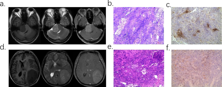
This multicenter, retrospective study included 313 meningioma patients, of which 70 were classified as high-grade (WHO II/III) and 243 as low-grade (WHO I). The Ki-67 expression was classified into low-expression (n = 216) and high-expression (n = 97) groups with a threshold of 5%. Among them, there were 128 patients with malignant biological behavior whose WHO grade or Ki-67 index increased either or both. Data from Center A and B are were utilized for model development, while data from Center C and D were used for external validation. Radiomic features were extracted from the maximum cross-sectional area (2D) region of Interest (ROI) and the whole tumor volume (3D) ROI using different paraments from the T1, T2-weighted, and T1 contrast-enhanced sequences (T1CE), followed by five independent feature selections and eight classifiers. 240 prediction models were constructed to predict the WHO grade, Ki-67 index and their combination respectively. Models were evaluated by cross-validation in training set (n = 224). Suitable models were chosen by comparing the cross-validation (CV) area under the curves (AUC) and their relative standard deviations (RSD). Clinical and radiological features were collected and analyzed; meaningful features were combined with radiomic features to establish the clinical-radiological-radiomic (CRR) models. The receiver operating characteristic (ROC) analysis was used to evaluate those models in validation set. Radiomic models and CRR models were compared by Delong test.
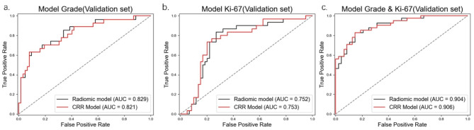
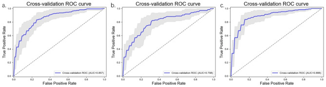
1218 and 1781 radiomic features were extracted from 2D ROI and 3D ROI of each sequence. The selected grade, Ki-67 index and their combination radiomic models were T1CE-2D-LASSO-LR, T1CE-3D-LASSO-NB, and T1CE-2D-LASSO-LR, with cross-validated AUCs on the training set were 0.857, 0.798, and 0.888, the RSDs were 0.06, 0.09, and 0.05, the validation set AUCs were 0.829, 0.752, and 0.904, respectively. Heterogeneous enhancement was found to be associated with high grade and Ki-67 status, while surrounding invasion was associated with the high grade status, peritumoral edema and cerebrospinal fluid space surrounding tumor were correlated with the high Ki-67 status. The Delong test showed that these significant radiological features did not significantly improve the predictive performance. The AUCs for CRR models predicting grade, Ki-67 index, and their combination in the validation set were 0.821, 0.753, and 0.906, respectively.
This study demonstrated that MRI-based machine learning models could effectively predict the grade, Ki-67 index of meningioma. Models considering these two indices might be valuable for improving the predictive sensitivity and comprehensiveness of prediction of malignant biological behavior of meningioma.
世界卫生组织(WHO)分级和 Ki-67 指数是评估脑膜瘤恶性生物学行为的独立指标。本研究旨在从 WHO 分级、Ki-67 指数及其组合的角度,利用基于 MRI 的机器学习模型预测脑膜瘤的恶性生物学行为。
这是一项多中心、回顾性研究,共纳入 313 例脑膜瘤患者,其中 70 例为高级别(WHO II/III 级),243 例为低级别(WHO I 级)。Ki-67 表达以 5%为界分为低表达(n=216)和高表达(n=97)组。其中,有 128 例患者的 WHO 分级或 Ki-67 指数升高。利用来自中心 A 和 B 的数据进行模型开发,同时利用来自中心 C 和 D 的数据进行外部验证。从 T1、T2 加权和 T1 对比增强序列(T1CE)的最大横截面积(2D)感兴趣区(ROI)和整个肿瘤体积(3D)ROI 中提取放射组学特征,然后使用不同参数进行五次独立特征选择和八种分类器。分别构建 240 个预测模型来预测 WHO 分级、Ki-67 指数及其组合。在训练集(n=224)中通过交叉验证评估模型。通过比较交叉验证(CV)曲线下面积(AUC)及其相对标准偏差(RSD)来选择合适的模型。收集和分析临床和影像学特征;将有意义的特征与放射组学特征相结合,建立临床-放射-放射组学(CRR)模型。利用受试者工作特征(ROC)分析在验证集上评估这些模型。通过 Delong 检验比较放射组学模型和 CRR 模型。
从每个序列的 2D ROI 和 3D ROI 中提取了 1218 个和 1781 个放射组学特征。选定的分级、Ki-67 指数及其组合的放射组学模型分别为 T1CE-2D-LASSO-LR、T1CE-3D-LASSO-NB 和 T1CE-2D-LASSO-LR,在训练集上的 CV AUC 分别为 0.857、0.798 和 0.888,RSD 分别为 0.06、0.09 和 0.05,验证集 AUC 分别为 0.829、0.752 和 0.904。不均匀强化与高级别和 Ki-67 状态相关,而周围侵犯与高级别相关,周围肿瘤水肿和肿瘤周围脑脊液空间与高 Ki-67 状态相关。Delong 检验表明这些显著的影像学特征并没有显著提高预测性能。CRR 模型在验证集上预测分级、Ki-67 指数及其组合的 AUC 分别为 0.821、0.753 和 0.906。
本研究表明,基于 MRI 的机器学习模型可以有效地预测脑膜瘤的分级、Ki-67 指数。考虑这些两个指数的模型可能有助于提高脑膜瘤恶性生物学行为预测的敏感性和全面性。