Department of Pathology, Botucatu Medical School, São Paulo State University, São Paulo, Brazil.
Division of Basic Science and Translational Research, Department of Obstetrics & Gynecology, The University of Texas Medical Branch at Galveston, Texas, USA.
Am J Reprod Immunol. 2023 Oct;90(4):e13770. doi: 10.1111/aji.13770.
Ascending bacterial infection is associated with ∼ 40% of spontaneous preterm birth (PTB), and Ureaplasma spp. is one of the most common bacteria isolated from the amniotic fluid. Developing novel in vitro models that mimic in vivo uterine physiology is essential to study microbial pathogenesis. We utilized the feto-maternal interface organ-on-chip (FMi-OOC) device and determined the propagation of Ureaplasma parvum, and its impact on cell signaling and inflammation.
FMi-OOC is a microphysiologic device mimicking fetal membrane/decidua interconnected through microchannels. The impact of resident decidual CD45 leukocytes was also determined by incorporating them into the decidual chamber in different combinations with U. parvum. We tested the propagation of live U. parvum from the decidual to the amniochorion membranes (immunocytochemistry and quantitative PCR), determined its impact on cytotoxicity (LDH assay), cell signaling (JESS Western Blot), cellular transition (immunostaining for vimentin and cytokeratin), and inflammation (cytokine bead array).
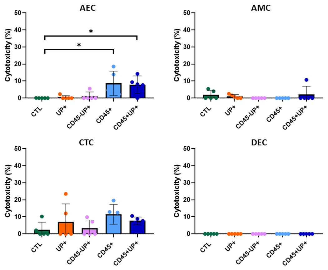
U. parvum transversed the chorion and reached the amnion epithelium after 72 hours but did not induce cell signaling kinases (p38MAPK and JNK) activation, or cellular transition (epithelial-mesenchymal), regardless of the presence of immune cells. The inflammatory response was limited to the choriodecidual interface and did not promote inflammation in the amnion layer.
Our data suggest that U. parvum is poorly immunogenic and does not produce massive inflammatory changes at the feto-maternal interface. We speculate that the presence of U. parvum may still compromise the feto-maternal interface making it susceptible to other pathogenic infection.
上行细菌感染与约 40%的自发性早产 (PTB) 有关,解脲支原体是从羊水分离出的最常见细菌之一。开发模拟体内子宫生理学的新型体外模型对于研究微生物发病机制至关重要。我们利用胎儿-母体界面器官芯片(FMi-OOC)设备,确定了微小脲原体的传播及其对细胞信号和炎症的影响。
FMi-OOC 是一种微生理设备,通过微通道模拟胎儿膜/蜕膜的相互连接。还通过将常驻蜕膜 CD45 白细胞以不同组合纳入蜕膜腔,确定了它们对微小脲原体的影响。我们测试了从蜕膜到羊膜绒毛膜的活微小脲原体的传播(免疫细胞化学和定量 PCR),确定了其对细胞毒性(LDH 测定)、细胞信号(JESS Western Blot)、细胞转化(波形蛋白和角蛋白免疫染色)和炎症(细胞因子珠阵列)的影响。
微小脲原体在 72 小时后穿过绒毛膜并到达羊膜上皮,但无论是否存在免疫细胞,都不会诱导细胞信号激酶(p38MAPK 和 JNK)的激活或细胞转化(上皮-间充质)。炎症反应仅限于绒毛膜-蜕膜界面,不会在羊膜层引起炎症。
我们的数据表明,微小脲原体免疫原性差,不会在胎儿-母体界面产生大量炎症变化。我们推测,微小脲原体的存在仍可能使胎儿-母体界面易受其他致病感染的影响。