Department of Ophthalmology, Renmin Hospital of Wuhan University, Wuhan, China.
Ann Med. 2023;55(2):2261494. doi: 10.1080/07853890.2023.2261494. Epub 2023 Sep 29.
To evaluate the retinal and choroidal microvasculature after one or two horizontal rectus muscle surgeries in strabismus patients using swept-source optical coherence tomography angiography (SS-OCTA).
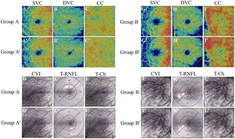
30 eyes of 26 patients who underwent horizontal rectus muscle surgery were included in this study. Group A, A' and Group B , B' respectively consisted preoperative and postoperative measurements of patients who underwent one or two horizontal rectus muscle surgeries. We analyzed the vessel density (VD) of the superficial vascular complex (SVC), the deep vascular complex (DVC), the choriocapillary layer (CC), choroidal vascular index (CVI), choroidal thickness (T-Ch) and retinal nerve fiber layer thickness (T-RNFL) preoperatively, and one week postoperatively.
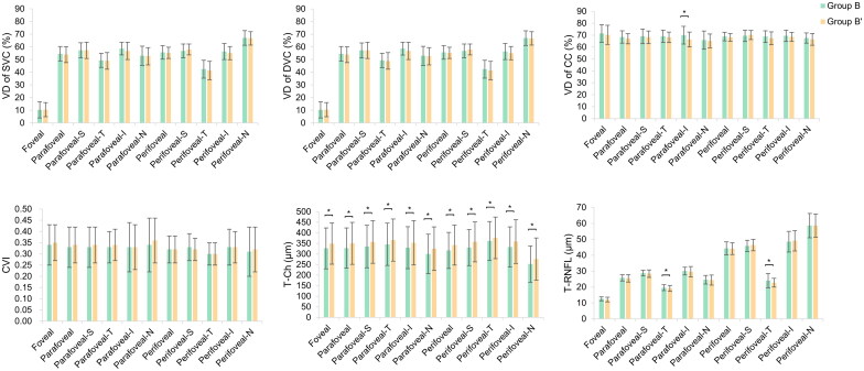
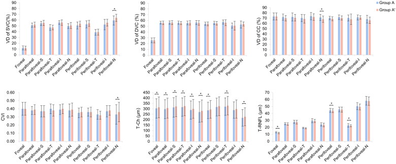
Only in the nasal sector of the perifoveal zone, the VD in SVC demonstrated a significant increase in Group A' ( = 0.027). There was a statistically significant difference in the VD changes of SVC between Group A and Group B ( = 0.043). The VD in DVC did not change significantly in the whole macular compared with the preoperative. Moreover, in both Group A' and Group B', the VD in CC showed a reduction in a single sector of the parafoveal area ( < 0.05). Group A' have increased CVI in the nasal sector of the perifoveal region ( = 0.008). In addition, the T-Ch increase in the perifoveal region was more significant in Group B' than in Group A' ( < 0.05). Group A' showed statistically significant decreases in T-RNFL in the foveal and parafoveal regions ( < 0.05).
This study revealed that the increase in choroidal thickness was more significant after two rectus muscle surgery. In addition, there were microvascular changes in sectional macular regions after strabismus surgery. OCTA is an excellent way to study the impact of strabismus surgery on the macular structure and blood flow.
使用扫频源光学相干断层血管造影术(SS-OCTA)评估斜视患者行一次或两次水平直肌手术后的视网膜和脉络膜微血管。
本研究纳入了 26 例斜视患者的 30 只眼,其中 A 组、A'组和 B 组、B'组分别为行一次或两次水平直肌手术后患者的术前和术后测量值。我们分析了术前和术后一周浅层血管丛(SVC)、深层血管丛(DVC)、脉络膜毛细血管层(CC)、脉络膜血管指数(CVI)、脉络膜厚度(T-Ch)和视网膜神经纤维层厚度(T-RNFL)的血管密度(VD)。
仅在中心凹旁鼻侧节段,A'组的 SVC 中 VD 显著增加(=0.027)。与 B 组相比,A 组 SVC 的 VD 变化有统计学差异(=0.043)。与术前相比,整个黄斑区的 DVC 中 VD 无明显变化。此外,在 A'组和 B'组中,CC 的 VD 在单一旁中心凹区节段减少(<0.05)。A'组在中心凹旁鼻侧区的 CVI 增加(=0.008)。此外,B'组的中心凹旁区 T-Ch 增加比 A'组更显著(<0.05)。A'组在中心凹和旁中心凹区的 T-RNFL 明显下降(<0.05)。
本研究表明,两次直肌手术后脉络膜厚度的增加更为显著。此外,斜视手术后节段性黄斑区存在微血管变化。OCTA 是研究斜视手术对黄斑结构和血流影响的极好方法。