The Spine and Spinal Cord Institute, Department of Neurosurgery, Gangnam Severance Hospital, Yonsei University College of Medicine, Seoul, 06273, Republic of Korea.
College of Medicine, Yonsei University Graduate School, Seoul, 03722, Republic of Korea.
Exp Mol Med. 2023 Oct;55(10):2190-2204. doi: 10.1038/s12276-023-01089-8. Epub 2023 Oct 2.
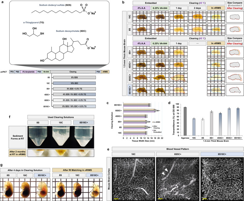
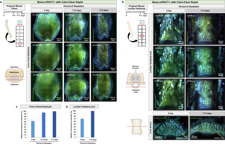
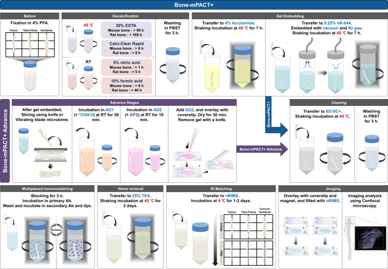
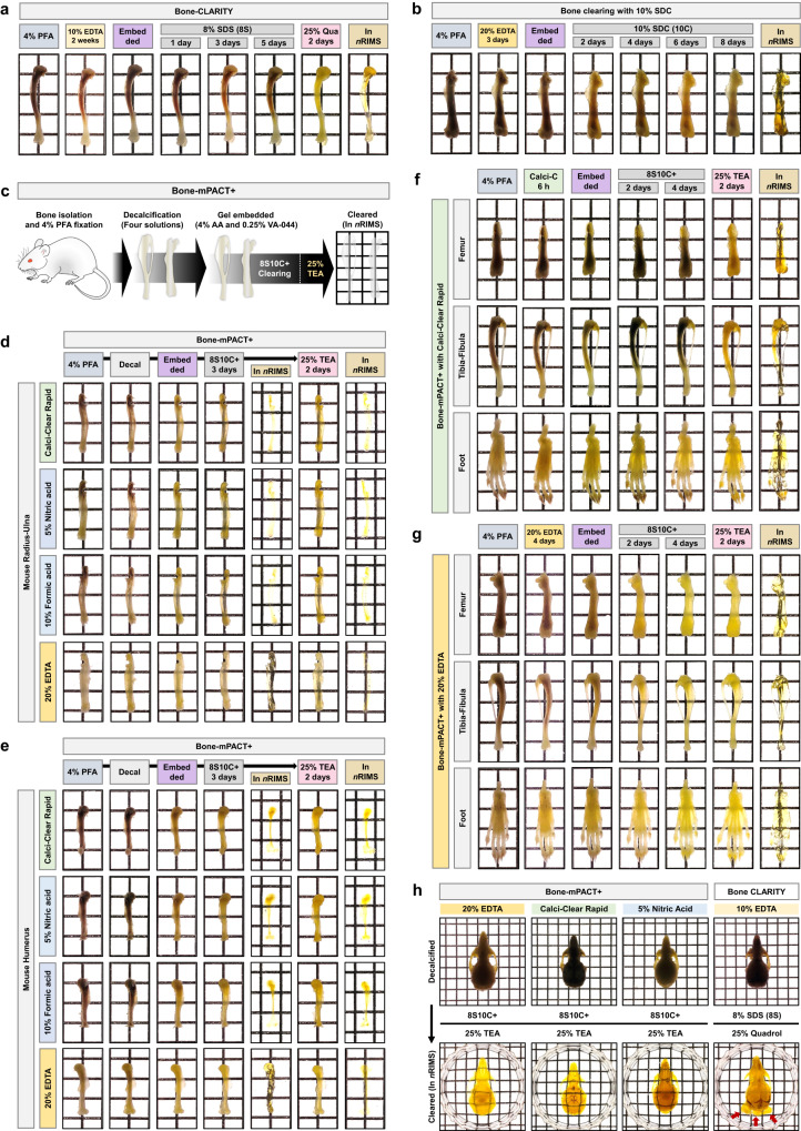
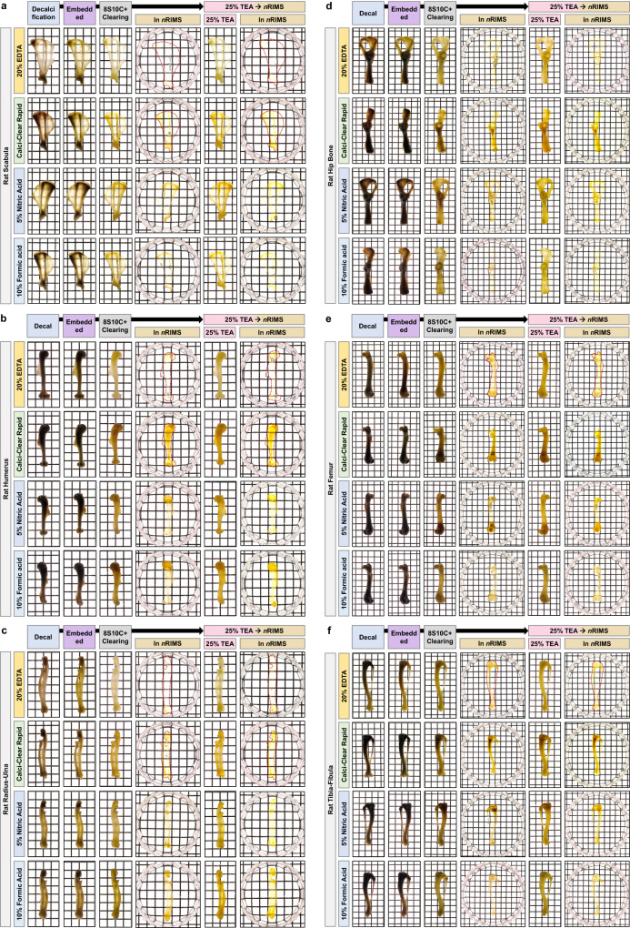
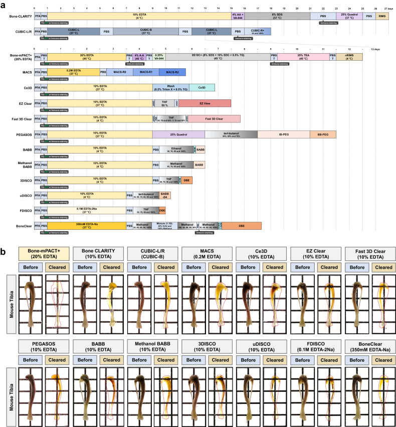
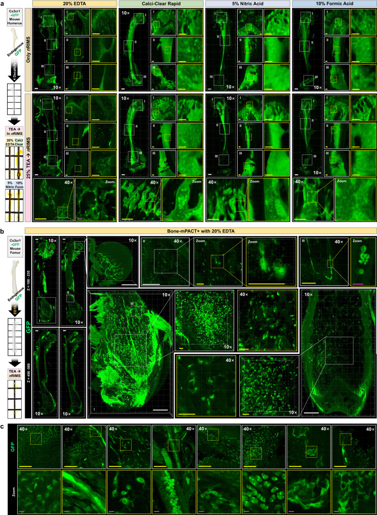
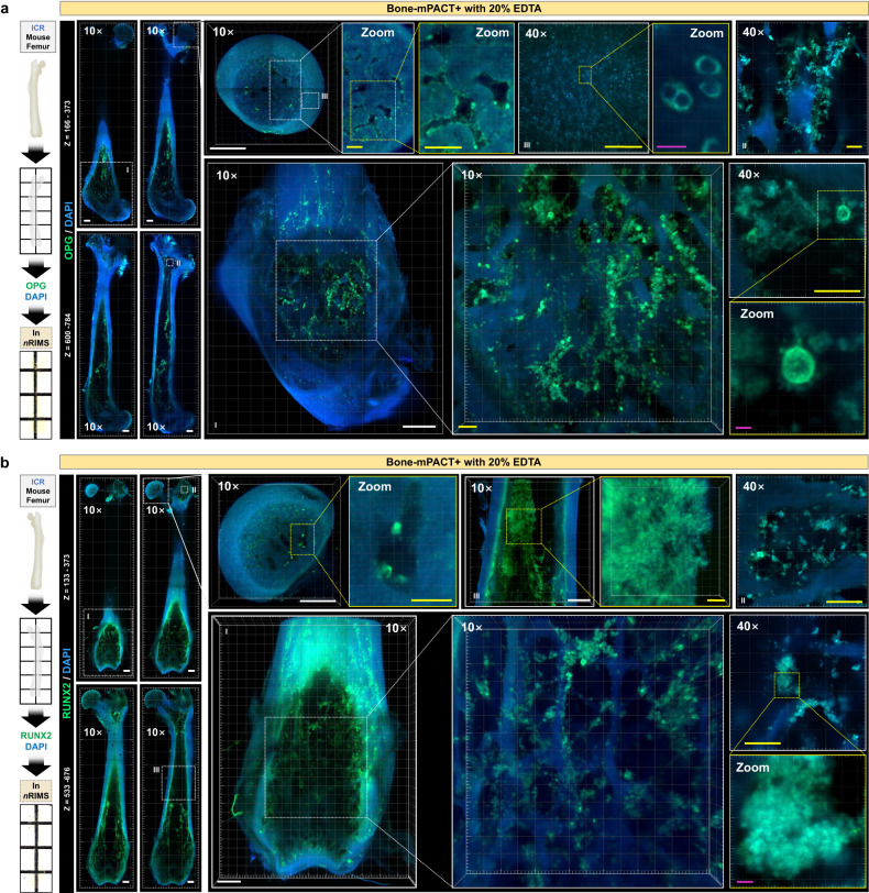
Recent developments in tissue clearing methods such as the passive clearing technique (PACT) have allowed three-dimensional analysis of biological structures in whole, intact tissues, thereby providing a greater understanding of spatial relationships and biological circuits. Nonetheless, the issues that remain in maintaining structural integrity and preventing tissue expansion/shrinkage with rapid clearing still inhibit the wide application of these techniques in hard bone tissues, such as femurs and tibias. Here, we present an optimized PACT-based bone-clearing method, Bone-mPACT+, that protects biological structures. Bone-mPACT+ and four different decalcifying procedures were tested for their ability to improve bone tissue clearing efficiency without sacrificing optical transparency; they rendered nearly all types of bone tissues transparent. Both mouse and rat bones were nearly transparent after the clearing process. We also present a further modification, the Bone-mPACT+ Advance protocol, which is specifically optimized for processing the largest and hardest rat bones for easy clearing and imaging using established tissue clearing methods.
近年来,组织透明化方法取得了一些进展,如被动透明化技术(PACT),可以对完整、未损伤的组织中的生物结构进行三维分析,从而更深入地了解空间关系和生物回路。然而,在快速透明化过程中保持结构完整性和防止组织扩张/收缩方面仍然存在一些问题,这限制了这些技术在股骨和胫骨等硬骨组织中的广泛应用。在这里,我们提出了一种优化的基于 PACT 的骨透明化方法 Bone-mPACT+,该方法可以保护生物结构。我们测试了 Bone-mPACT+和四种不同的脱钙程序,以确定它们在不牺牲光学透明度的情况下提高骨组织透明化效率的能力;它们使几乎所有类型的骨组织都变得透明。在透明化处理后,老鼠和大鼠的骨骼几乎都是透明的。我们还提出了进一步的改进,即 Bone-mPACT+ Advance 方案,该方案专门针对最大和最硬的大鼠骨骼进行了优化,以便使用现有的组织透明化方法轻松进行透明化和成像。