From the University of Lyon, INSA-Lyon, Claude Bernard Lyon 1 University, UJM-Saint Etienne, CNRS, Inserm, Villeurbanne, France (P.C.D., L.B., S.A.S.M.); Department of Cardiovascular and Thoracic Radiology, Louis Pradel Hospital, Hospices Civils de Lyon, 59 Boulevard Pinel, 69500 Bron, France (P.C.D., S.B., L.B., S.A.S.M.); Claude Bernard Lyon 1 University, Villeurbanne, France (S.B.); Department of Radiology and Nuclear Medicine, Erasmus Medical Center, Rotterdam, the Netherlands (E.H.G.O., R.P.J.B.); Department of Radiology, University of Pennsylvania, Philadelphia, Pa (D.P.C.); Department of Radiology and Imaging Sciences, Emory University, Atlanta, Ga (A.P.); Department of Biomedical Engineering, Georgia Institute of Technology, Atlanta, Ga (A.P.); and Winship Cancer Institute, Atlanta, Ga (A.P.).
Radiology. 2023 Oct;309(1):e222432. doi: 10.1148/radiol.222432.
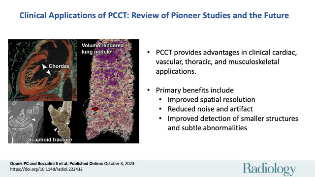
CT systems equipped with photon-counting detectors (PCDs), referred to as photon-counting CT (PCCT), are beginning to change imaging in several subspecialties, such as cardiac, vascular, thoracic, and musculoskeletal radiology. Evidence has been building in the literature underpinning the many advantages of PCCT for different clinical applications. These benefits derive from the distinct features of PCDs, which are made of semiconductor materials capable of converting photons directly into electric signal. PCCT advancements include, among the most important, improved spatial resolution, noise reduction, and spectral properties. PCCT spatial resolution on the order of 0.25 mm allows for the improved visualization of small structures (eg, small vessels, arterial walls, distal bronchi, and bone trabeculations) and their pathologies, as well as the identification of previously undetectable anomalies. In addition, blooming artifacts from calcifications, stents, and other dense structures are reduced. The benefits of the spectral capabilities of PCCT are broad and include reducing radiation and contrast material dose for patients. In addition, multiple types of information can be extracted from a single data set (ie, multiparametric imaging), including quantitative data often regarded as surrogates of functional information (eg, lung perfusion). PCCT also allows for a novel type of CT imaging, K-edge imaging. This technique, combined with new contrast materials specifically designed for this modality, opens the door to new applications for imaging in the future.
配备光子计数探测器(PCD)的 CT 系统,称为光子计数 CT(PCCT),开始改变心脏、血管、胸部和肌肉骨骼放射学等多个亚专业的成像方式。越来越多的文献证据支持 PCCT 在不同临床应用中的许多优势。这些优势源于 PCD 的独特特性,它由能够将光子直接转换为电信号的半导体材料制成。PCCT 的进展包括最重要的改进,如空间分辨率、噪声降低和光谱特性。PCCT 的空间分辨率约为 0.25 毫米,可改善对小结构(如小血管、动脉壁、远端支气管和骨小梁)及其病变的可视化,以及识别以前无法检测到的异常。此外,还减少了来自钙化、支架和其他高密度结构的辉光伪影。PCCT 的光谱功能的优势广泛,包括降低患者的辐射和对比剂剂量。此外,还可以从单个数据集提取多种类型的信息(即多参数成像),包括通常被视为功能信息替代物的定量数据(例如,肺灌注)。PCCT 还允许进行一种新型的 CT 成像,即 K 边成像。这项技术与专为该模式设计的新型对比材料相结合,为未来的成像应用开辟了新的途径。