Gupta Himanshu, Spanopoulous Basil, Lubat Edward, Krinsky Glenn, Rutledge John, Fortier Jacqueline H, Grau Juan, Tayal Rajiv
The Valley Hospital, Ridgewood, NJ, USA.
The University of Ottawa Heart Institute, Ottawa, Ontario, Canada.
Heliyon. 2023 Sep 9;9(9):e19974. doi: 10.1016/j.heliyon.2023.e19974. eCollection 2023 Sep.
Recent guidelines provide broader support for the use of less invasive imaging modalities for the evaluation of patients with stable chest pain. Coronary CT angiography (CCTA) uses increasingly sophisticated techniques to improve evaluation of coronary lesions. The purpose of this study is to describe one center's experience implementing AI-assisted advanced imaging techniques to diagnose coronary artery disease.
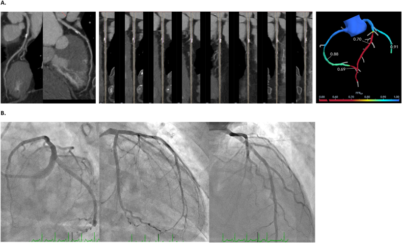
MATERIALS & METHODS: Retrospective study of patients who had AI-assisted CCTA interpretation, including a subgroup who underwent fractional flow reserve CT (FFR-CT) and invasive coronary angiography. Descriptive statistics summarized baseline characteristics and univariate statistics compared findings between groups of patients with and without anatomically and hemodynamically significant lesions based on FFR-CT. For patients who underwent invasive coronary angiography, concordance between CCTA and angiography was evaluated.
Of 532 included patients, AI-assisted CCTA identified statistically significant difference in calcification scores, plaque types and total plaque volume between lesions <50% and ≥50% stenosis. CCTA results were mostly concordant with invasive coronary angiography. Importantly, we identified a subset of patients with less than 50% anatomical stenosis that demonstrated physiologically significant stenosis on FFR-CT and invasive coronary angiography.
AI-assisted CCTA and other advanced techniques are a tool to support high quality diagnostic assessment of coronary lesions in a clinical environment. Combined CCTA with FFRCT in mild to moderate coronary stenosis identifies patients with hemodynamically significant stenosis even when quantitative stenosis is <50%. Implementation of AI-assisted coronary CT angiography is feasible in a community hospital setting, but these technologies do not replace the need for expert review and clinical correlation.
近期指南为使用侵入性较小的成像方式评估稳定型胸痛患者提供了更广泛的支持。冠状动脉CT血管造影(CCTA)采用日益复杂的技术来改善对冠状动脉病变的评估。本研究的目的是描述一个中心实施人工智能辅助先进成像技术诊断冠状动脉疾病的经验。
对接受人工智能辅助CCTA解读的患者进行回顾性研究,包括一个接受血流储备分数CT(FFR-CT)和有创冠状动脉造影的亚组。描述性统计总结了基线特征,单变量统计比较了基于FFR-CT有或无解剖学和血流动力学显著病变的患者组之间的结果。对于接受有创冠状动脉造影的患者,评估CCTA与造影之间的一致性。
在纳入的532例患者中,人工智能辅助CCTA发现狭窄<50%和≥50%的病变在钙化评分、斑块类型和总斑块体积方面存在统计学显著差异。CCTA结果与有创冠状动脉造影大多一致。重要的是,我们确定了一部分解剖学狭窄小于50%的患者,其在FFR-CT和有创冠状动脉造影上显示出生理学上显著的狭窄。
人工智能辅助CCTA和其他先进技术是在临床环境中支持对冠状动脉病变进行高质量诊断评估的工具。在轻度至中度冠状动脉狭窄中,将CCTA与FFRCT相结合可识别出血流动力学显著狭窄的患者,即使定量狭窄<50%。在社区医院环境中实施人工智能辅助冠状动脉CT血管造影是可行的,但这些技术并不能取代专家审查和临床关联的需求。