Chen Yi, You Ningning, Yang Chaoyu, Zhang Jinshun
Departments of Gastroenterology, Taizhou Hospital of Zhejiang Province Affiliated to Wenzhou Medical University, Taizhou, China.
Health Management Center, Taizhou Hospital of Zhejiang Province Affiliated to Wenzhou Medical University, Taizhou, China.
Heliyon. 2023 Sep 9;9(9):e20037. doi: 10.1016/j.heliyon.2023.e20037. eCollection 2023 Sep.
Infection with () may increase atherosclerosis, which can lead to carotid plaque formation. Our study examined the relationship between infection and carotid plaque formation, and its underlying mechanisms.
A total of 36,470 people who underwent physical examination in Taizhou Hospital Health Examination Center from June 2017 to June 2022 were included in this study. All people participated in the urease test, neck ultrasound, blood pressure detection, anthropometric measurement and biochemical laboratory examination. In addition, the GSE27411 and GSE28829 datasets in the Gene Expression Omnibus (GEO) database were used to analyze the mechanism of infection and atherosclerosis progression.
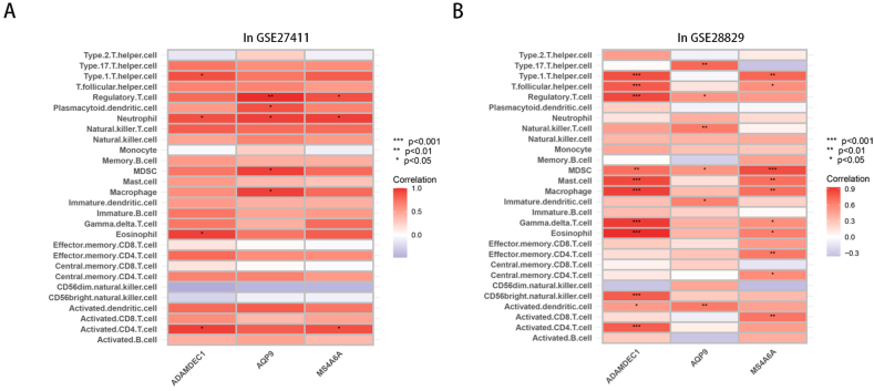
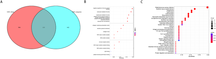
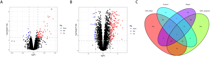
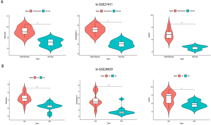
infection, sex, age, blood lipids, blood pressure, fasting blood glucose, glycated hemoglobin and body mass index were risk factors for carotid plaque formation. An independent risk factor was still evident in the multivariate logistic regression analysis, indicating infection. Furthermore, after weighted gene coexpression network analysis (WGCNA), we discovered 555 genes linked to both infection and the advancement of atherosclerosis. Gene Ontology (GO) and Kyoto Encyclopedia of Genes and Genomes (KEGG) enrichment analyses revealed a strong correlation between these genes and immunity, infection, and immune disorders. SsGSEA analysis showed that infection and atherosclerosis included changes in the immune microenvironment. Finally, three genes MS4A6A, ADAMDEC1 and AQP9 were identified to be involved in the formation of atherosclerosis after infection. Conclusion: Our research affirms that is a unique contributor to the formation of carotid plaque, examines the immune microenvironment associated with infection and advanced carotid atherosclerosis, and offers fresh perspectives on how infection leads to atherosclerosis.
感染()可能会增加动脉粥样硬化,进而导致颈动脉斑块形成。我们的研究探讨了感染与颈动脉斑块形成之间的关系及其潜在机制。
本研究纳入了2017年6月至2022年6月在台州医院健康体检中心接受体检的36470人。所有人均参与了尿素酶检测、颈部超声检查、血压检测、人体测量和生化实验室检查。此外,利用基因表达综合数据库(GEO)中的GSE27411和GSE28829数据集分析感染与动脉粥样硬化进展的机制。
感染、性别、年龄、血脂、血压、空腹血糖、糖化血红蛋白和体重指数是颈动脉斑块形成的危险因素。多因素逻辑回归分析中仍有一个独立危险因素,即感染。此外,经过加权基因共表达网络分析(WGCNA),我们发现了555个与感染和动脉粥样硬化进展均相关的基因。基因本体论(GO)和京都基因与基因组百科全书(KEGG)富集分析显示这些基因与免疫、感染和免疫紊乱密切相关。单样本基因集富集分析(SsGSEA)表明感染和动脉粥样硬化包括免疫微环境的变化。最后,确定了三个基因MS4A6A、ADAMDEC1和AQP9参与感染后动脉粥样硬化的形成。结论:我们的研究证实是颈动脉斑块形成的独特促成因素,研究了与感染和晚期颈动脉粥样硬化相关的免疫微环境,并为感染导致动脉粥样硬化的机制提供了新的视角。