Department of Radiology, General Hospital of Northern Theater Command, 83 Wenhua Road, Shenyang, 110016, Liaoning Province, China.
BMC Cardiovasc Disord. 2023 Oct 10;23(1):500. doi: 10.1186/s12872-023-03541-z.
The purpose of this study was to explore the relationship between quantitative epicardial adipose tissue (EAT) based on coronary computed tomography angiography (CCTA) and coronary slow flow (CSF).
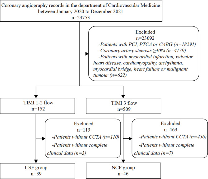
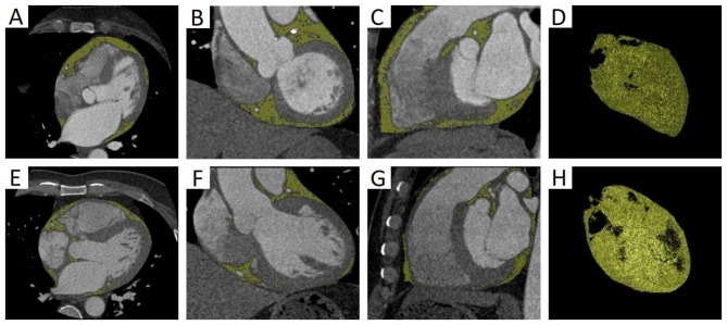
A total of 85 patients with < 40% coronary stenosis on diagnostic coronary angiography were included in this retrospective study between January 2020 and December 2021. A semi-automatic method was developed for EAT quantification on CCTA images. According to the thrombolysis in myocardial infarction flow grade, the patients were divided into CSF group (n = 39) and normal coronary flow group (n = 46). Multivariate logistic regression was used to explore the relationship between EAT and CSF. Receiver operating characteristic (ROC) curve was plotted to evaluate the diagnostic value of EAT in CSF.
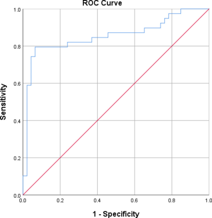
EAT volume in the CSF group was significantly higher than that of the normal coronary flow group (128.83± 21.59 mL vs. 101.87± 18.56 mL, P < 0.001). There was no significant difference in epicardial fat attenuation index between the two groups (P > 0.05). Multivariate logistic regression analysis showed that EAT volume was independently related to CSF [odds ratio (OR) = 4.82, 95% confidence interval (CI): 3.06-7.27, P < 0.001]. The area under ROC curve for EAT volume in identifying CSF was 0.86 (95% CI: 0.77-0.95). The optimal cutoff value of 118.46 mL yielded a sensitivity of 0.80 and a specificity of 0.94.
Increased EAT volume based on CCTA is strongly associated with CSF. This preliminary finding paves the way for future and larger studies aimed to definitively recognize the diagnostic value of EAT in CSF.
本研究旨在探讨基于冠状动脉计算机断层扫描血管造影(CCTA)的定量心外膜脂肪组织(EAT)与冠状动脉慢血流(CSF)之间的关系。
本回顾性研究纳入了 2020 年 1 月至 2021 年 12 月期间在诊断性冠状动脉造影中存在<40%冠状动脉狭窄的 85 例患者。采用半自动方法对 CCTA 图像进行 EAT 定量。根据心肌梗死溶栓血流分级,将患者分为 CSF 组(n=39)和正常冠状动脉血流组(n=46)。采用多变量逻辑回归探讨 EAT 与 CSF 的关系。绘制受试者工作特征(ROC)曲线评估 EAT 在 CSF 中的诊断价值。
CSF 组的 EAT 体积明显高于正常冠状动脉血流组(128.83±21.59ml 比 101.87±18.56ml,P<0.001)。两组心外膜脂肪衰减指数无显著差异(P>0.05)。多变量逻辑回归分析显示,EAT 体积与 CSF 独立相关[比值比(OR)=4.82,95%置信区间(CI):3.06-7.27,P<0.001]。EAT 体积用于识别 CSF 的 ROC 曲线下面积为 0.86(95%CI:0.77-0.95)。最佳截断值为 118.46ml 时,敏感性为 0.80,特异性为 0.94。
基于 CCTA 的 EAT 体积增加与 CSF 密切相关。这一初步发现为未来更大规模的研究奠定了基础,旨在明确 EAT 在 CSF 中的诊断价值。