Department of Oral and Maxillofacial Radiology, School of Dentistry, Shiraz University of Medical Sciences, Shiraz, Iran.
School of Dentistry, Shiraz University of Medical Sciences, Shiraz, Iran.
BMC Oral Health. 2023 Oct 13;23(1):753. doi: 10.1186/s12903-023-03440-x.
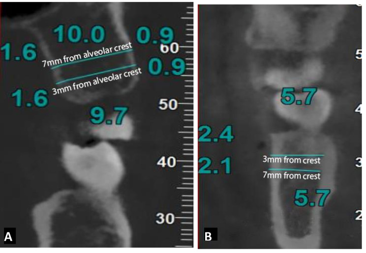
The purpose of this study was to evaluate the effect of the density and the thickness of the cortical and the cancellous bone at selected inter-radicular areas in subjects with different facial growth patterns using cone beam computed tomography (CBCT) in order to choose the optimal area for miniscrew insertion.
From 150 CBCT scans, 45 scans were included in the study. The subjects were categorized into three groups based on their skeletal growth pattern according to SN-GoMe angle and facial height index. Cortical and cancellous bone density and thickness were measured at the selected inter-radicular areas.
Compared to the other two groups, the hyperdivergent group had thinner cortical bone in the anterior region of the maxilla between the central and the lateral incisors on the buccal side at 4 mm from the alveolar crest (P-value: 0.012) and on the palatal side at 7 mm from the alveolar crest (P-value: 0.030). Cancellous bone density values in these areas were higher in subjects with hypodivergent and hyperdivergent growth pattern. Furthermore, in hyperdivergent group less dense cortical bone in the posterior region of the maxilla on the palatal side between the second premolar and the first molar (p-value: 0.020) and on the buccal side between the first molar and the second molar (p-value: 0.038 & 0.047) was observed. No significant differences were found in the mandible between the three groups. No significant differences were found between the male and the female subjects.
Hyperdivegents presented thinner cortical bone in the anterior of the maxilla between the central and the lateral incisors. Less dense cortical bone was found between maxillary second premolar and first molar on the palatal side and also between the maxillary first molar and the second molar on the buccal side in this group too. Normal showed higher density values in the posterior of the maxilla compared to the other two groups. No significant differences were found among three groups in mandible.
本研究旨在使用锥形束 CT(CBCT)评估不同面部生长模式受试者在选定的根间区域的皮质骨和松质骨的密度和厚度,以选择植入微螺钉的最佳区域。
从 150 例 CBCT 扫描中,选取 45 例纳入研究。根据 SN-GoMe 角和面部高度指数,将受试者分为三组。在选定的根间区域测量皮质骨和松质骨的密度和厚度。
与其他两组相比,高角组在上颌前区中切牙和侧切牙之间的颊侧牙槽嵴 4mm 处(P 值:0.012)和腭侧牙槽嵴 7mm 处(P 值:0.030)皮质骨较薄。低角组和高角组的这些区域的松质骨密度值较高。此外,在高角组中,上颌后区第二前磨牙和第一磨牙之间的腭侧(P 值:0.020)和颊侧(P 值:0.038 和 0.047)皮质骨密度较低。下颌骨在三组之间无显著差异。男女之间也没有发现显著差异。
高角组在上颌中切牙和侧切牙之间的前区皮质骨较薄。在上颌第二前磨牙和第一磨牙之间的腭侧以及上颌第一磨牙和第二磨牙之间的颊侧也发现皮质骨密度较低。与其他两组相比,上颌后区的正常组的密度值较高。下颌骨在三组之间无显著差异。