Department of Surgery, University of California San Diego, La Jolla, California; Department of Surgery, VA San Diego Healthcare System, La Jolla, California.
Department of Surgical Oncology, City of Hope National Medical Center, Duarte, California.
J Surg Res. 2024 Jan;293:701-708. doi: 10.1016/j.jss.2023.08.038. Epub 2023 Oct 14.
Gastric cancer poses a major therapeutic challenge. Improved visualization of tumor margins at the time of gastrectomy with fluorescent tumor-specific antibodies could improve outcomes. The present report demonstrates the potential of targeting gastric cancer with a humanized anti-carcinoembryonic antigen (CEA) antibody in orthotopic mouse models.
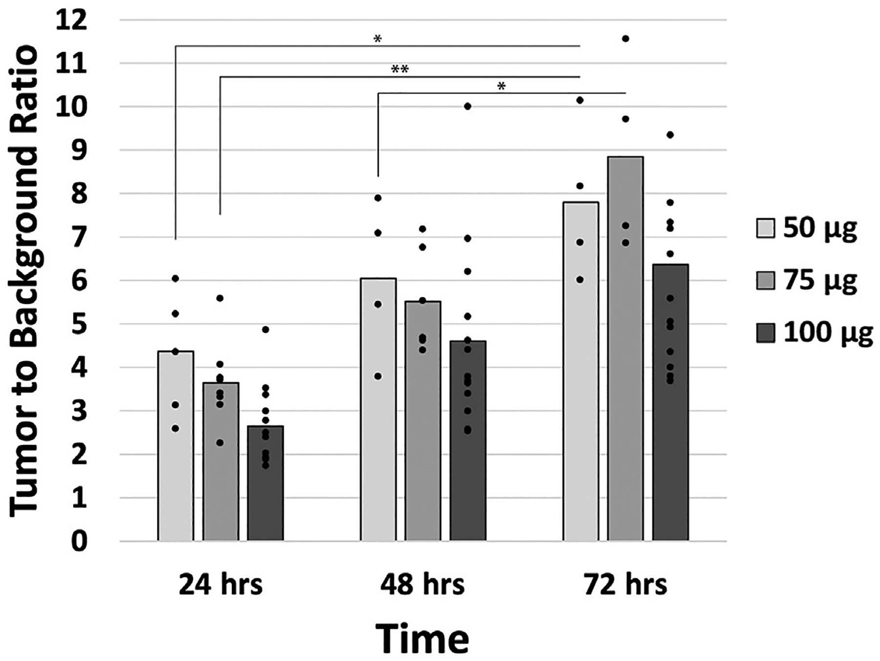
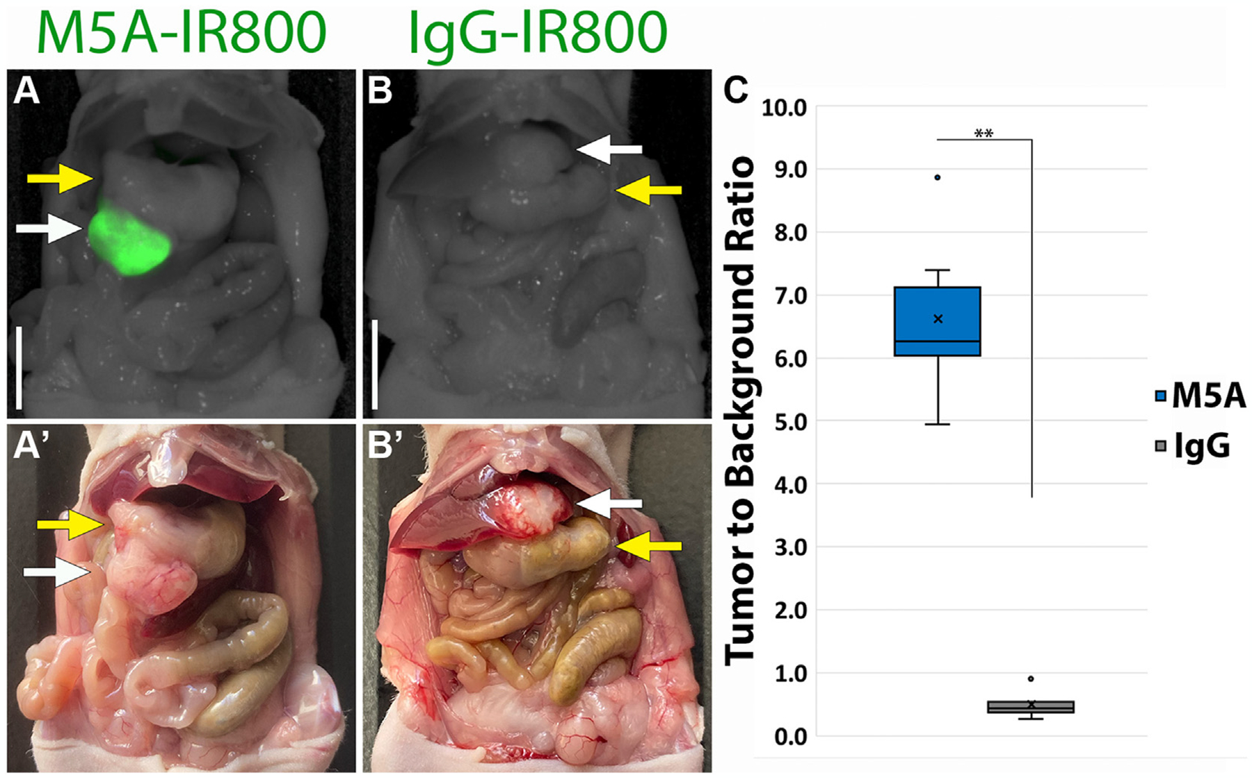
MKN45 cells were injected subcutaneously into nude mice to establish xenograft models. Tumor fragments collected from subcutaneous models were then implanted into the greater curvature of the stomach to establish orthotopic models. For tumor labeling, a humanized anti-CEA antibody (M5A) and IgG as a control, were conjugated with the near-infrared dye IRDye800CW. Time (24-72 h) and dose (50-100 μg) response curves were performed in subcutaneous models. Orthotopic models received 50 μg of M5A-IR800 or 50 μg IgG-IR800 as a control and were imaged after 72 h. Fluorescence imaging was performed on the mice using the LI-COR Pearl Imaging System.
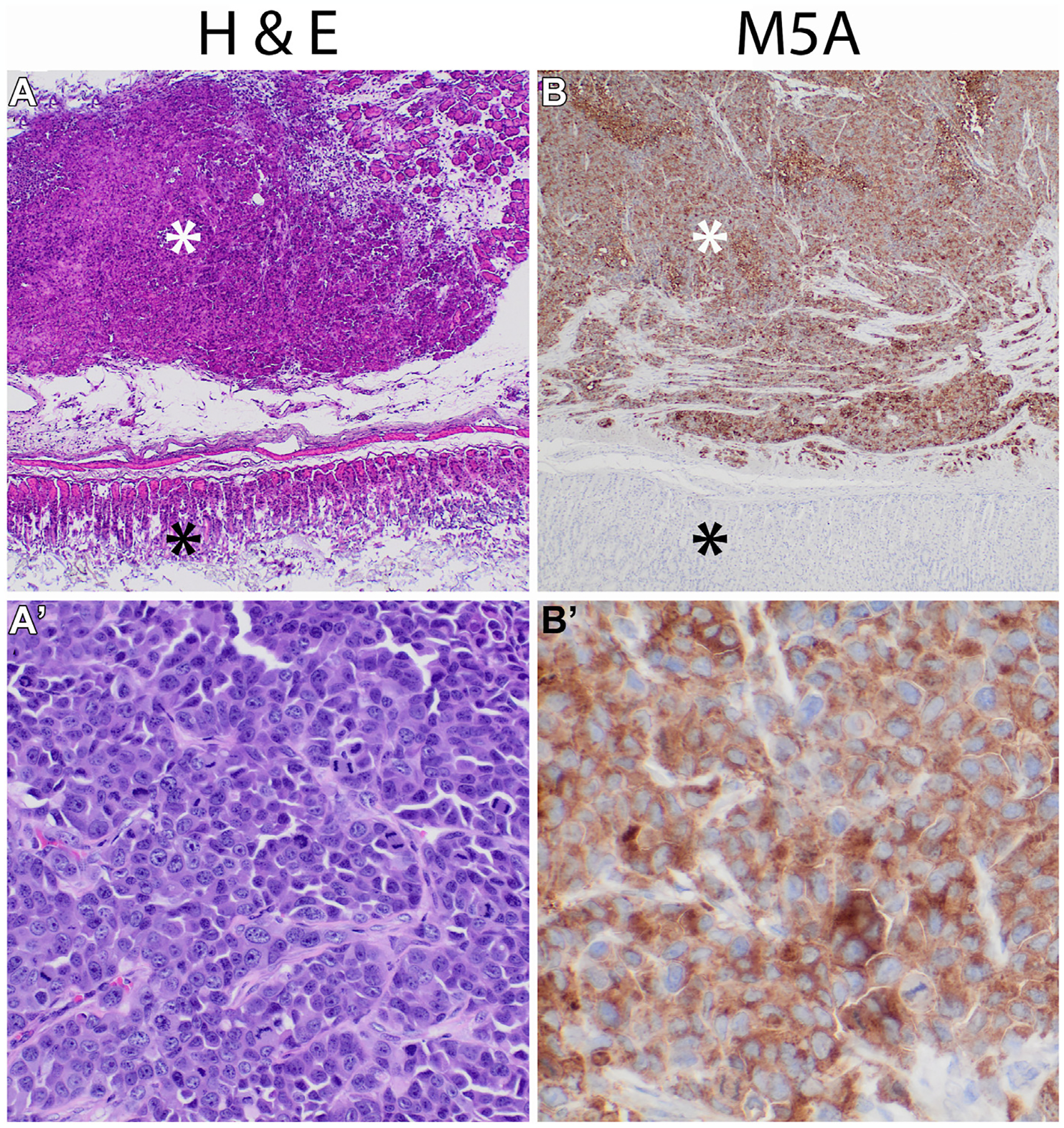
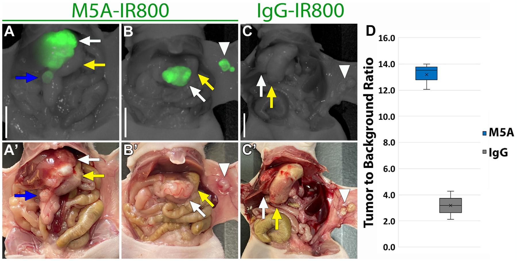
In subcutaneous models, tumor to background ratios (TBRs) reached 8.85 at 72 h. Median TBRs of orthotopic model primary tumors were 6.25 (interquartile range [IQR] 6.03-7.12) for M5A-IR800 compared to 0.42 (IQR 0.38-0.54) for control. Abdominal wall metastasis median TBRs were 13.52 (IQR 12.79-13.76) for M5A-IR800 and 3.19 (IQR 2.65-3.73) for the control. Immunohistochemistry confirmed CEA expression within tumors.
Humanized anti-CEA antibodies conjugated to near-infrared dyes provide specific labeling of gastric cancers in mouse models. Orthotopic models demonstrated bright and specific labeling with TBRs greater than ten times that of control. This tumor-specific fluorescent antibody is a promising potential clinical tool for improving visualization of gastric cancer margins at time of surgical resection.
胃癌是一个治疗方面的重大挑战。在胃切除术中使用荧光肿瘤特异性抗体提高肿瘤边缘的可视性可能会改善结果。本报告展示了使用针对癌胚抗原(CEA)的人源化抗体靶向胃癌在原位小鼠模型中的潜力。
将 MKN45 细胞皮下注射到裸鼠中建立异种移植模型。从皮下模型中采集的肿瘤组织碎片然后植入胃大弯处建立原位模型。为了进行肿瘤标记,将人源化抗 CEA 抗体(M5A)和 IgG 作为对照与近红外染料 IRDye800CW 偶联。在皮下模型中进行时间(24-72 小时)和剂量(50-100μg)的反应曲线。原位模型接受 50μg 的 M5A-IR800 或 50μg IgG-IR800 作为对照,并在 72 小时后进行成像。使用 LI-COR Pearl 成像系统对小鼠进行荧光成像。
在皮下模型中,肿瘤与背景的比值(TBR)在 72 小时达到 8.85。M5A-IR800 治疗的原位模型原发性肿瘤的中位数 TBR 为 6.25(四分位距[IQR]6.03-7.12),而对照组为 0.42(IQR 0.38-0.54)。腹壁转移的中位数 TBR 为 M5A-IR800 为 13.52(IQR 12.79-13.76),对照组为 3.19(IQR 2.65-3.73)。免疫组织化学证实肿瘤内存在 CEA 表达。
与人源化 CEA 抗体偶联的近红外染料可特异性标记小鼠模型中的胃癌。与对照组相比,原位模型的 TBR 高 10 倍以上,具有明亮而特异性的标记。这种肿瘤特异性荧光抗体是一种很有前途的临床工具,可提高手术切除时胃癌边缘的可视性。