Sadeghi Fatemeh, Sheikhzadeh Peyman, Farzanehfar Saeed, Ghafarian Pardis, Moafpurian Yalda, Ay Mohammadreza
Department of Medical Physics and Biomedical Engineering, Tehran University of Medical Sciences, Tehran, Iran.
Research Center for Molecular and Cellular Imaging (RCMCI), Advanced Medical Technologies and Equipment Institute (AMTEI), Tehran University of Medical Sciences, Tehran, Iran.
EJNMMI Phys. 2023 Oct 16;10(1):63. doi: 10.1186/s40658-023-00587-y.
The Q.Clear algorithm is a fully convergent iterative image reconstruction technique. We hypothesize that different PET/CT scanners with distinct crystal properties will require different optimal settings for the Q.Clear algorithm. Many studies have investigated the improvement of the Q.Clear reconstruction algorithm on PET/CT scanner with LYSO crystals and SiPM detectors. We propose an optimum penalization factor (β) for the detection of rectal cancer and its metastases using a BGO-based detector PET/CT system which obtained via accurate and comprehensive phantom and clinical studies.
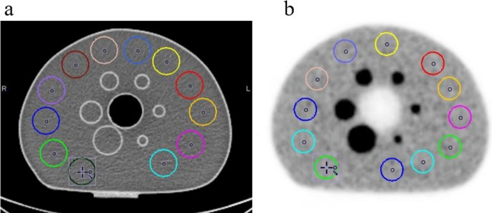
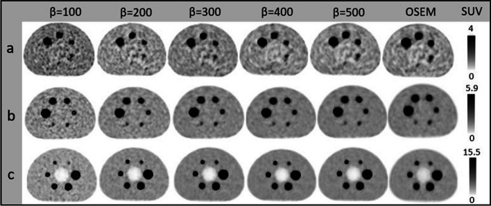
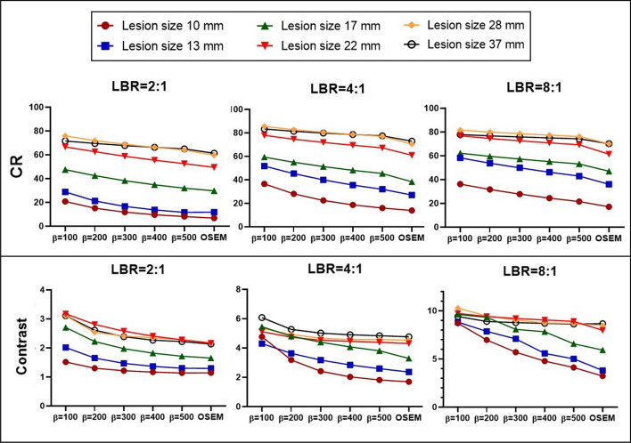
F-FDG PET-CT scans were acquired from NEMA phantom with lesion-to-background ratio (LBR) of 2:1, 4:1, 8:1, and 15 patients with rectal cancer. Clinical lesions were classified into two size groups. OSEM and Q.Clear (β value of 100-500) reconstruction was applied. In Q.Clear, background variability (BV), contrast recovery (CR), signal-to-noise ratio (SNR), SUVmax, and signal-to-background ratio (SBR) were evaluated and compared to OSEM.
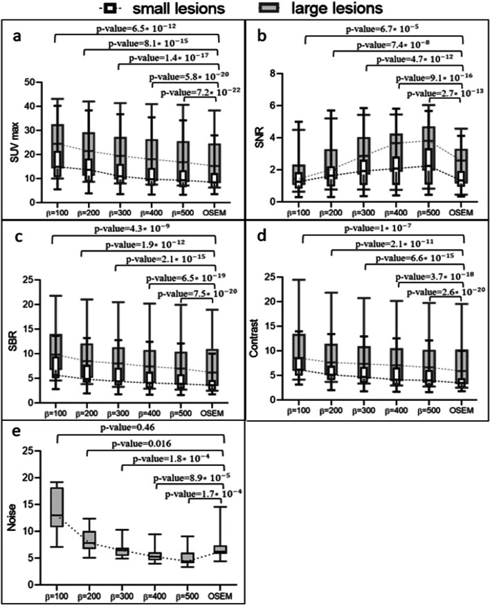
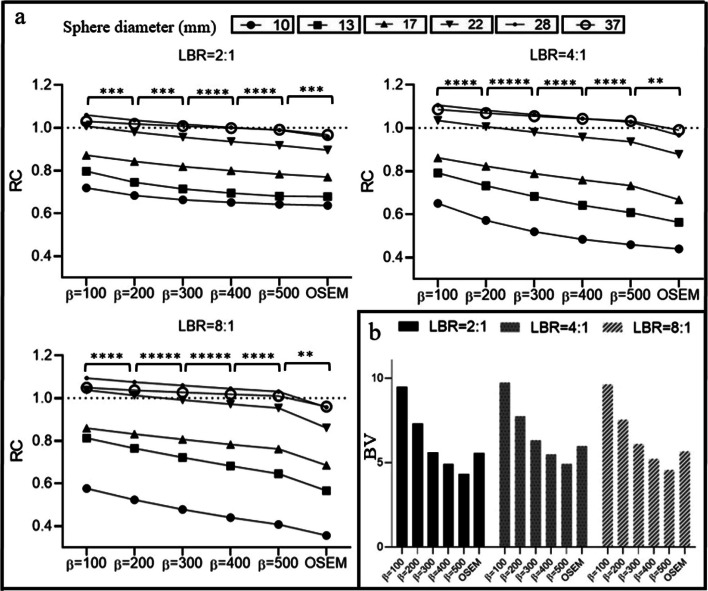
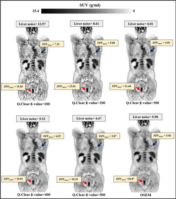
OSEM had 11.5-18.6% higher BV than Q.Clear using β value of 500. Conversely, RC from OSEM to Q.Clear using β value of 500 decreased by 3.3-7.7% for a sphere with a diameter of 10 mm and 2.5-5.1% for a sphere with a diameter of 37 mm. Furthermore, the increment of contrast using a β value of 500 was 5.2-8.1% in the smallest spheres compared to OSEM. When the β value was increased from 100 to 500, the SNR increased by 49.1% and 30.8% in the smallest and largest spheres at LBR 2:1, respectively. At LBR of 8:1, the relative difference of SNR between β value of 100 and 500 was 43.7% and 44.0% in the smallest and largest spheres, respectively. In the clinical study, as β increased from 100 to 500, the SUVmax decreased by 47.7% in small and 31.1% in large lesions. OSEM demonstrated the least SUVmax, SBR, and contrast. The decrement of SBR and contrast using OSEM were 13.6% and 12.9% in small and 4.2% and 3.4%, respectively, in large lesions.
Implementing Q.Clear enhances quantitative accuracies through a fully convergent voxel-based image approach, employing a penalization factor. In the BGO-based scanner, the optimal β value for small lesions ranges from 200 for LBR 2:1 to 300 for LBR 8:1. For large lesions, the optimal β value is between 400 for LBR 2:1 and 500 for LBR 8:1. We recommended β value of 300 for small lesions and β value of 500 for large lesions in clinical study.
Q.Clear算法是一种完全收敛的迭代图像重建技术。我们假设,具有不同晶体特性的不同PET/CT扫描仪对Q.Clear算法需要不同的最佳设置。许多研究已经探讨了Q.Clear重建算法在配备LYSO晶体和硅光电倍增管(SiPM)探测器的PET/CT扫描仪上的改进情况。我们通过精确且全面的体模和临床研究,为使用基于BGO探测器的PET/CT系统检测直肠癌及其转移灶提出了一个最佳惩罚因子(β)。
从NEMA体模获取了病变与背景比值(LBR)为2:1、4:1、8:1的F-FDG PET-CT扫描图像,并对15例直肠癌患者进行了扫描。临床病变被分为两个大小组。应用有序子集期望最大化(OSEM)和Q.Clear(β值为100 - 500)重建。在Q.Clear中,评估背景变异性(BV)、对比度恢复(CR)、信噪比(SNR)、最大标准摄取值(SUVmax)和信号与背景比值(SBR),并与OSEM进行比较。
使用β值为500时,OSEM的BV比Q.Clear高11.5 - 18.6%。相反,对于直径为10 mm的球体,使用β值为500时,从OSEM到Q.Clear的对比度恢复(CR)下降了3.3 - 7.7%,对于直径为37 mm的球体下降了2.5 - 5.1%。此外,与OSEM相比,使用β值为500时,最小球体的对比度增量为5.2 - 8.1%。当β值从100增加到500时,在LBR为2:1时,最小和最大球体的SNR分别增加了49.1%和30.8%。在LBR为8:1时,β值为100和500之间,最小和最大球体的SNR相对差异分别为43.7%和44.0%。在临床研究中,随着β值从100增加到500,小病变的SUVmax下降了47.7%,大病变下降了31.1%。OSEM的SUVmax、SBR和对比度最低。使用OSEM时,小病变的SBR和对比度下降分别为13.6%和12.9%,大病变分别为4.2%和3.4%。
通过采用惩罚因子的基于体素的完全收敛图像方法实施Q.Clear可提高定量准确性。在基于BGO的扫描仪中,小病变的最佳β值范围为LBR 2:1时为200至LBR 8:1时为300。对于大病变,最佳β值在LBR 2:1时为400至LBR 8:1时为500之间。我们建议在临床研究中,小病变的β值为300,大病变的β值为500。