Sung Jonghoo, Barratt Kate R, Pederson Stephen M, Chenu Chantal, Reichert Ines, Atkins Gerald J, Anderson Paul H, Smitham Peter J
Centre for Orthopaedic and Trauma Research, Adelaide Medical School, Faculty of Health and Medical Sciences, The University of Adelaide, Adelaide, Australia.
Clinical and Health Sciences, University of South Australia, Adelaide, Australia.
Bone Joint Res. 2023 Oct 17;12(10):657-666. doi: 10.1302/2046-3758.1210.BJR-2023-0062.R1.
Impaired fracture repair in patients with type 2 diabetes mellitus (T2DM) is not fully understood. In this study, we aimed to characterize the local changes in gene expression (GE) associated with diabetic fracture. We used an unbiased approach to compare GE in the fracture callus of Zucker diabetic fatty (ZDF) rats relative to wild-type (WT) littermates at three weeks following femoral osteotomy.
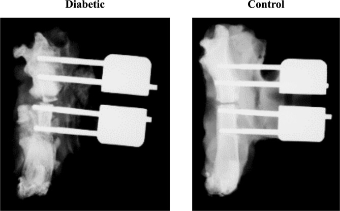
Zucker rats, WT and homozygous for leptin receptor mutation (ZDF), were fed a moderately high-fat diet to induce T2DM only in the ZDF animals. At ten weeks of age, open femoral fractures were simulated using a unilateral osteotomy stabilized with an external fixator. At three weeks post-surgery, the fractured femur from each animal was retrieved for analysis. Callus formation and the extent of healing were assessed by radiograph and histology. Bone tissue was processed for total RNA extraction and messenger RNA (mRNA) sequencing (mRNA-Seq).
Radiographs and histology demonstrated impaired fracture healing in ZDF rats with incomplete bony bridge formation and an influx of intramedullary inflammatory tissue. In comparison, near-complete bridging between cortices was observed in Sham WT animals. Of 13,160 genes, mRNA-Seq analysis identified 13 that were differentially expressed in ZDF rat callus, using a false discovery rate (FDR) threshold of 10%. Seven genes were upregulated with high confidence (FDR = 0.05) in ZDF fracture callus, most with known roles in inflammation.
These findings suggest that elevated or prolonged inflammation contributes to delayed fracture healing in T2DM. The identified genes may be used as biomarkers to monitor and treat delayed fracture healing in diabetic patients.
2型糖尿病(T2DM)患者骨折修复受损的情况尚未完全明确。在本研究中,我们旨在描述与糖尿病性骨折相关的基因表达(GE)的局部变化。我们采用无偏倚方法,比较股骨截骨术后三周时,Zucker糖尿病肥胖(ZDF)大鼠与野生型(WT)同窝仔鼠骨折痂中的GE。
Zucker大鼠,野生型和瘦素受体突变纯合子(ZDF),喂食适度高脂肪饮食,仅在ZDF动物中诱导T2DM。在10周龄时,使用外固定器固定的单侧截骨术模拟开放性股骨骨折。术后三周,取出每只动物的骨折股骨进行分析。通过X线片和组织学评估骨痂形成和愈合程度。对骨组织进行处理以提取总RNA并进行信使核糖核酸(mRNA)测序(mRNA-Seq)。
X线片和组织学显示,ZDF大鼠骨折愈合受损,骨桥形成不完全,髓内炎性组织浸润。相比之下,在假手术WT动物中观察到皮质之间几乎完全桥接。在13160个基因中,mRNA-Seq分析使用10%的错误发现率(FDR)阈值,鉴定出13个在ZDF大鼠骨痂中差异表达的基因。在ZDF骨折痂中,7个基因高度上调(FDR = 0.05),大多数在炎症中具有已知作用。
这些发现表明,炎症升高或持续时间延长导致T2DM患者骨折愈合延迟。所鉴定的基因可作为生物标志物,用于监测和治疗糖尿病患者的延迟骨折愈合。