Pereira M, Gohin S, Lund N, Hvid A, Smitham P J, Oddy M J, Reichert I, Farlay D, Roux J P, Cleasby M E, Chenu C
Department of Comparative Biomedical sciences, Royal Veterinary College, Royal College Street, London, NW1 0TU, UK.
University College London, London, UK.
Osteoporos Int. 2017 Jan;28(1):309-320. doi: 10.1007/s00198-016-3718-0. Epub 2016 Jul 28.
In contrast to previously reported elevations in serum sclerostin levels in diabetic patients, the present study shows that the impaired bone microarchitecture and cellular turnover associated with type 2 diabetes mellitus (T2DM)-like conditions in ZDF rats are not correlated with changes in serum and bone sclerostin expression.
T2DM is associated with impaired skeletal structure and a higher prevalence of bone fractures. Sclerostin, a negative regulator of bone formation, is elevated in serum of diabetic patients. We aimed to relate changes in bone architecture and cellular activities to sclerostin production in the Zucker diabetic fatty (ZDF) rat.
Bone density and architecture were measured by micro-CT and bone remodelling by histomorphometry in tibiae and femurs of 14-week-old male ZDF rats and lean Zucker controls (n = 6/group).
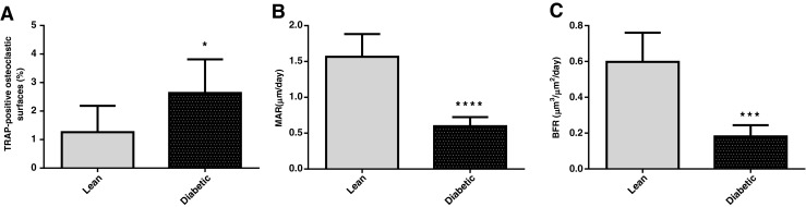
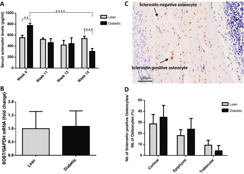
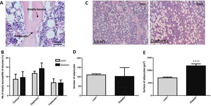
ZDF rats showed lower trabecular bone mineral density and bone mass compared to controls, due to decreases in bone volume and thickness, along with impaired bone connectivity and cortical bone geometry. Bone remodelling was impaired in diabetic rats, demonstrated by decreased bone formation rate and increased percentage of tartrate-resistant acid phosphatase-positive osteoclastic surfaces. Serum sclerostin levels (ELISA) were higher in ZDF compared to lean rats at 9 weeks (+40 %, p < 0.01), but this difference disappeared as their glucose control deteriorated and by week 14, ZDF rats had lower sclerostin levels than control rats (-44 %, p < 0.0001). Bone sclerostin mRNA (qPCR) and protein (immunohistochemistry) were similar in ZDF, and lean rats at 14 weeks and genotype did not affect the number of empty osteocytic lacunae in cortical and trabecular bone.
T2DM results in impaired skeletal architecture through altered remodelling pathways, but despite altered serum levels, it does not appear that sclerostin contributes to the deleterious effect of T2DM in rat bone.
与先前报道的糖尿病患者血清硬化蛋白水平升高相反,本研究表明,ZDF大鼠中与2型糖尿病(T2DM)样病症相关的骨微结构受损和细胞更新与血清及骨硬化蛋白表达的变化无关。
T2DM与骨骼结构受损和骨折患病率较高有关。硬化蛋白是骨形成的负调节因子,在糖尿病患者血清中升高。我们旨在将Zucker糖尿病脂肪(ZDF)大鼠的骨结构和细胞活性变化与硬化蛋白产生联系起来。
通过显微CT测量14周龄雄性ZDF大鼠和瘦型Zucker对照大鼠(每组n = 6)胫骨和股骨的骨密度和结构,并通过组织形态计量学测量骨重塑。
与对照组相比,ZDF大鼠的小梁骨矿物质密度和骨量较低,这是由于骨体积和厚度减小,以及骨连接性和皮质骨几何形状受损所致。糖尿病大鼠的骨重塑受损,表现为骨形成率降低和抗酒石酸酸性磷酸酶阳性破骨细胞表面百分比增加。在9周时,ZDF大鼠的血清硬化蛋白水平(ELISA)高于瘦大鼠(升高40%,p < 0.01),但随着血糖控制恶化,这种差异消失,到14周时,ZDF大鼠的硬化蛋白水平低于对照大鼠(降低44%,p < 0.0001)。14周时,ZDF大鼠和瘦大鼠的骨硬化蛋白mRNA(qPCR)和蛋白质(免疫组织化学)相似,基因型不影响皮质骨和小梁骨中空骨陷窝的数量。
T2DM通过改变重塑途径导致骨骼结构受损,但尽管血清水平改变,硬化蛋白似乎并未促成T2DM对大鼠骨骼的有害作用。