Wacinski Piotr, Madejczyk Andrzej, Kondracki Bartosz, O'Kane Peter, Wacinski Jakub, Kijewski Bartosz, Kawiak Andrzej, Binko Paweł, Głowniak Andrzej, Wysokiński Andrzej
Department of Cardiology, Medical University of Lublin, SPSK4 Hospital, Lublin, Poland.
Dorset Heart Centre, The Royal Bournemouth Hospital, Castle Lane East, Bournemouth, Dorset, UK.
Postepy Kardiol Interwencyjnej. 2023 Sep;19(3):209-216. doi: 10.5114/aic.2023.131473. Epub 2023 Sep 27.
Complex, coronary stenosis remains a technical challenge that may be responsible for in-stent restenosis and vessel thrombosis. Here we investigated the efficacy and safety of excimer laser coronary atherectomy (ELCA) with contrast mix injection for improving vessel wall stent apposition in undilatable, mostly calcified lesions.
To assess ELCA with contrast mix injection in complex, stented, calcified coronary lesions.
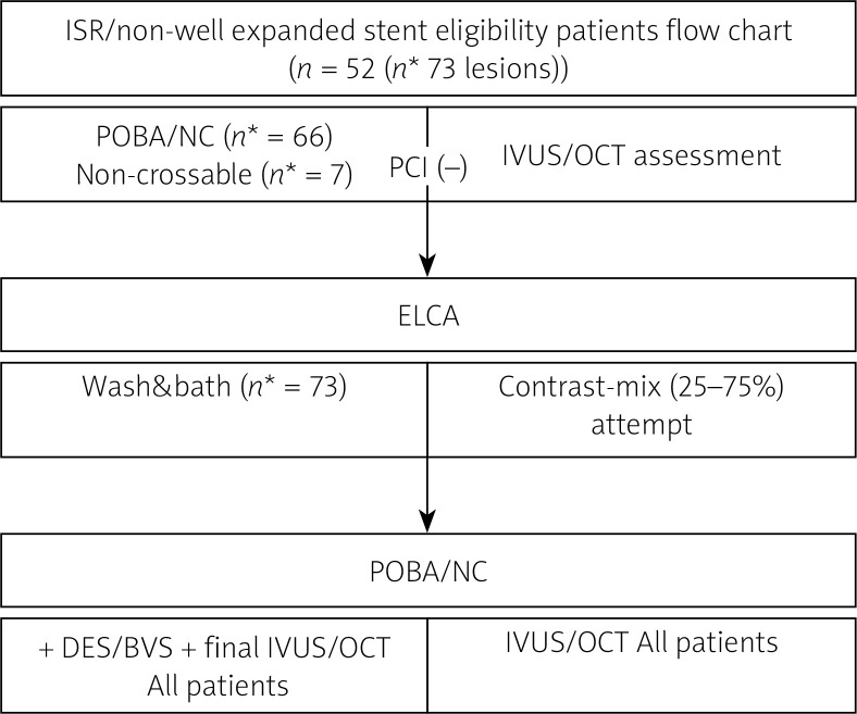
This prospective single-center observational study enrolled 52 consecutive patients (73 lesions), with suboptimal stents implanted in de novo lesions and lesions requiring in-stent restenosis (ISR) due to stent underexpansion using all available means to achieve an optimal result. Patients presenting with ST-segment elevation myocardial infarction were excluded. All patients underwent coronary angiography 6 months after ELCA with intravascular ultrasound or optical coherence tomography study. We used contrast media mixed with saline (25-75%) to supply maximum laser energy output when a standard approach was unsuccessful. Procedural success was defined as relative stent expansion of > 80% minimal stent area (MSA) divided by average reference lumen area.
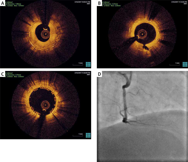
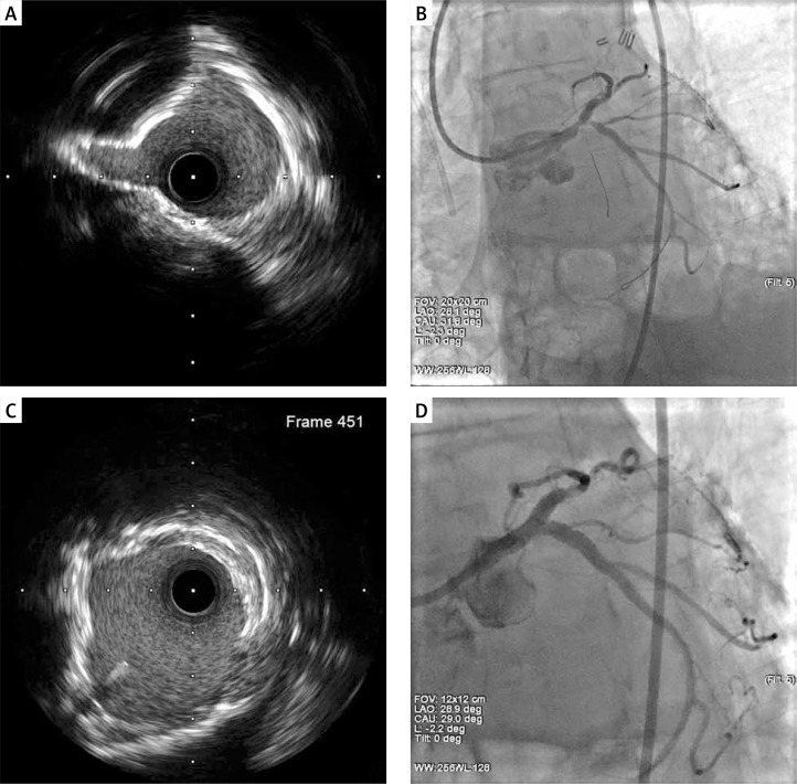
Procedural success was achieved in all cases. The cross-sectional area measured in treated segment improved significantly from 2.9 (0.72) mm to 7.3 (0.79) mm after ELCA. The in-hospital device-oriented major adverse cardiac event (DOCE) rate was 9.6%. No vessel perforation occurred during ELCA. After 6 months, the DOCE rate was 13.4%, while the rate of target lesion revascularization (TLR) was 8.2%.
This registry confirms the efficacy and safety of ELCA with contrast mix injection as a possible approach for stent expansion/ISR in failed PCI.
复杂的冠状动脉狭窄仍然是一项技术挑战,可能导致支架内再狭窄和血管血栓形成。在此,我们研究了准分子激光冠状动脉斑块旋切术(ELCA)联合造影剂混合注射在改善不可扩张、主要为钙化病变的血管壁支架贴壁方面的疗效和安全性。
评估准分子激光冠状动脉斑块旋切术联合造影剂混合注射在复杂的、置入支架的钙化冠状动脉病变中的效果。
这项前瞻性单中心观察性研究连续纳入了52例患者(73处病变),这些患者的初发病变植入了效果欠佳的支架,以及因支架扩张不足而需要进行支架内再狭窄(ISR)治疗的病变,采用所有可用方法以获得最佳结果。排除出现ST段抬高型心肌梗死的患者。所有患者在ELCA术后6个月接受冠状动脉造影,并进行血管内超声或光学相干断层扫描研究。当标准方法不成功时,我们使用与盐水混合(25 - 75%)的造影剂来提供最大激光能量输出。手术成功定义为相对支架扩张率>80%,即最小支架面积(MSA)除以平均参考管腔面积。
所有病例均取得手术成功。ELCA术后,治疗节段的横截面积从2.9(0.72)mm显著改善至7.3(0.79)mm。院内器械相关主要不良心脏事件(DOCE)发生率为9.6%。ELCA期间未发生血管穿孔。6个月后,DOCE发生率为13.4%,而靶病变血运重建(TLR)率为8.2%。
该注册研究证实了准分子激光冠状动脉斑块旋切术联合造影剂混合注射作为PCI失败时支架扩张/ISR的一种可能方法的疗效和安全性。