• 文献检索
  • 文档翻译
  • 深度研究
  • 学术资讯
  • Suppr Zotero 插件Zotero 插件
  • 邀请有礼
  • 套餐&价格
  • 历史记录
应用&插件
Suppr Zotero 插件Zotero 插件浏览器插件Mac 客户端Windows 客户端微信小程序
定价
高级版会员购买积分包购买API积分包
服务
文献检索文档翻译深度研究API 文档MCP 服务
关于我们
关于 Suppr公司介绍联系我们用户协议隐私条款
关注我们

Suppr 超能文献

核心技术专利:CN118964589B侵权必究
粤ICP备2023148730 号-1Suppr @ 2026

文献检索

告别复杂PubMed语法,用中文像聊天一样搜索,搜遍4000万医学文献。AI智能推荐,让科研检索更轻松。

立即免费搜索

文件翻译

保留排版,准确专业,支持PDF/Word/PPT等文件格式,支持 12+语言互译。

免费翻译文档

深度研究

AI帮你快速写综述,25分钟生成高质量综述,智能提取关键信息,辅助科研写作。

立即免费体验

CT上高衰减肺部异常的逐步诊断算法

Stepwise diagnostic algorithm for high-attenuation pulmonary abnormalities on CT.

作者信息

Fukuda Taiki, Egashira Ryoko, Ueno Midori, Hashisako Mikiko, Sumikawa Hiromitsu, Tominaga Junya, Yamada Daisuke, Fukuoka Junya, Misumi Shigeki, Ojiri Hiroya, Hatabu Hiroto, Johkoh Takeshi

机构信息

Department of Radiology, The Jikei University School of Medicine, 3-25-8, Nishi-Shimbashi, Minato-Ku, Tokyo, 105-8461, Japan.

Department of Radiology, Faculty of Medicine, Saga University, 5-1-1, Nabeshima, Saga-City, Saga, 849-8501, Japan.

出版信息

Insights Imaging. 2023 Oct 20;14(1):177. doi: 10.1186/s13244-023-01501-x.

DOI:10.1186/s13244-023-01501-x
PMID:37857741
原文链接:https://pmc.ncbi.nlm.nih.gov/articles/PMC10587054/
Abstract

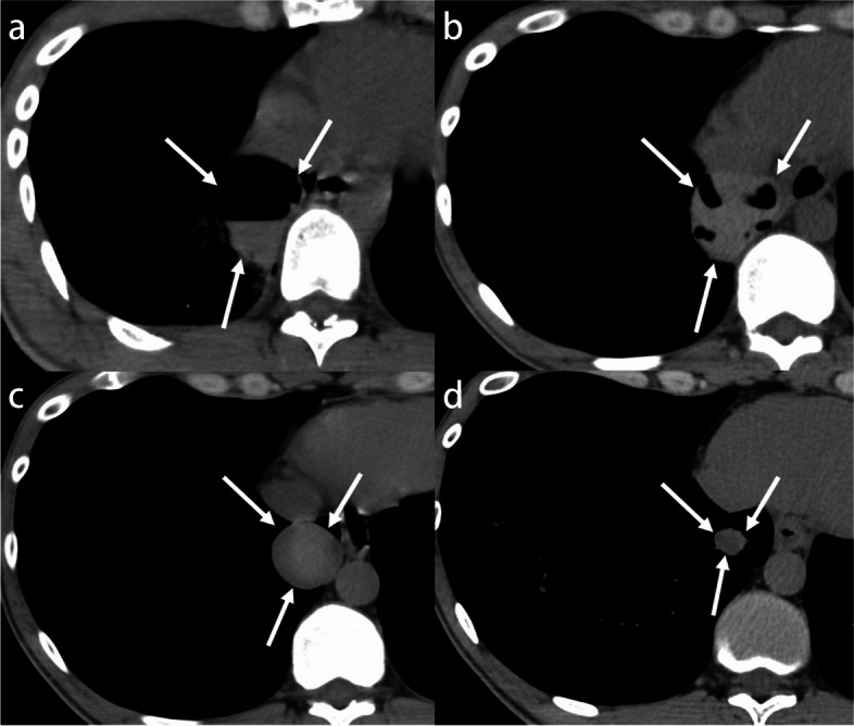
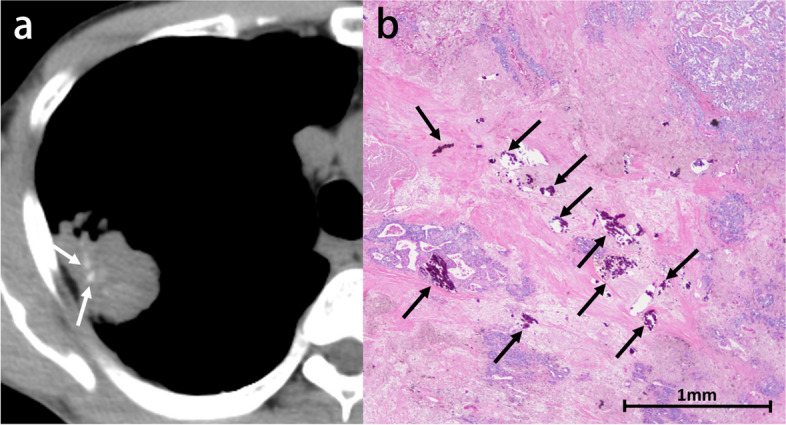
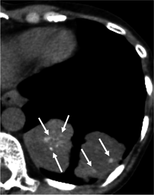
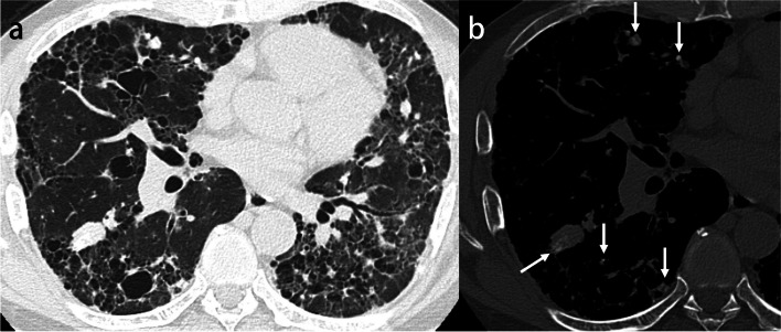
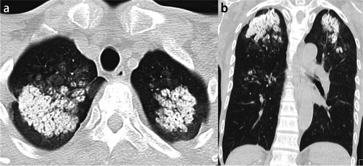
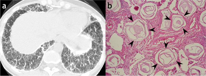
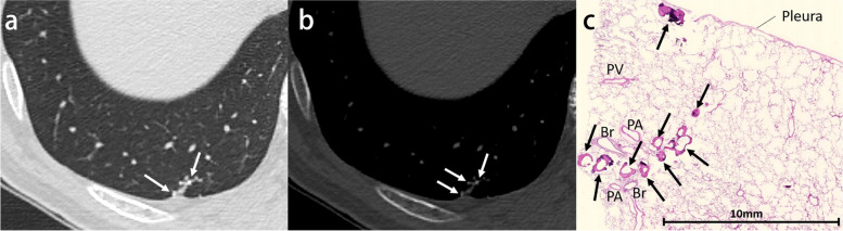
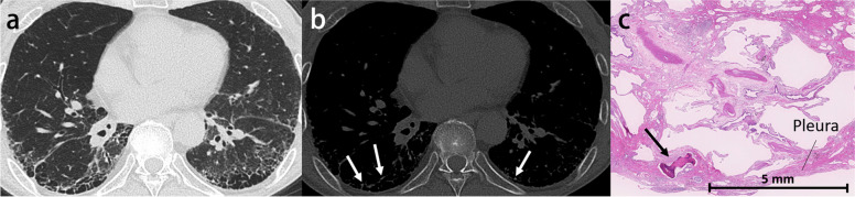
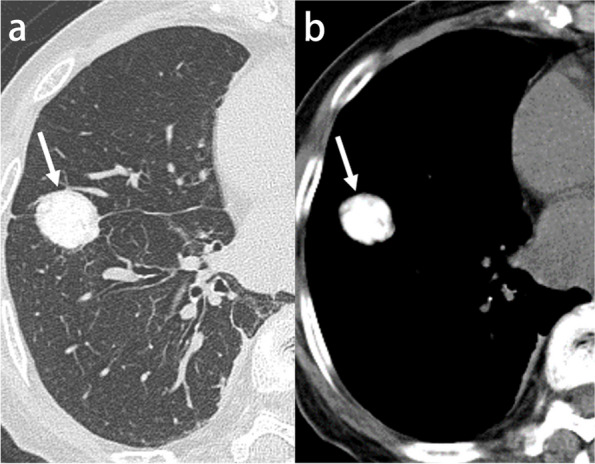
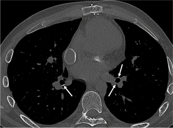
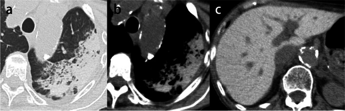
High-attenuation pulmonary abnormalities are commonly seen on CT. These findings are increasingly encountered with the growing number of CT examinations and the wide availability of thin-slice images. The abnormalities include benign lesions, such as infectious granulomatous diseases and metabolic diseases, and malignant tumors, such as lung cancers and metastatic tumors. Due to the wide spectrum of diseases, the proper diagnosis of high-attenuation abnormalities can be challenging. The assessment of these abnormal findings requires scrutiny, and the treatment is imperative. Our proposed stepwise diagnostic algorithm consists of five steps. Step 1: Establish the presence or absence of metallic artifacts. Step 2: Identify associated nodular or mass-like soft tissue components. Step 3: Establish the presence of solitary or multiple lesions if identified in Step 2. Step 4: Ascertain the predominant distribution in the upper or lower lungs if not identified in Step 2. Step 5: Identify the morphological pattern, such as linear, consolidation, nodular, or micronodular if not identified in Step 4. These five steps to diagnosing high-attenuation abnormalities subdivide the lesions into nine categories. This stepwise radiologic diagnostic approach could help to narrow the differential diagnosis for various pulmonary high-attenuation abnormalities and to achieve a precise diagnosis.Critical relevance statement Our proposed stepwise diagnostic algorithm for high-attenuation pulmonary abnormalities may help to recognize a variety of those high-attenuation findings, to determine whether the associated diseases require further investigation, and to guide appropriate patient management. Key points • To provide a stepwise diagnostic approach to high-attenuation pulmonary abnormalities.• To familiarize radiologists with the varying cause of high-attenuation pulmonary abnormalities.• To recognize which high-attenuation abnormalities require scrutiny and prompt treatment.

摘要

高衰减肺部异常在CT上很常见。随着CT检查数量的增加和薄层图像的广泛应用,这些发现越来越多地被遇到。这些异常包括良性病变,如感染性肉芽肿疾病和代谢性疾病,以及恶性肿瘤,如肺癌和转移性肿瘤。由于疾病谱广泛,对高衰减异常进行正确诊断可能具有挑战性。对这些异常发现的评估需要仔细审查,且治疗势在必行。我们提出的逐步诊断算法包括五个步骤。步骤1:确定是否存在金属伪影。步骤2:识别相关的结节状或肿块样软组织成分。步骤3:如果在步骤2中识别出病变,确定是单发还是多发。步骤4:如果在步骤2中未识别出病变,确定在上肺还是下肺的主要分布。步骤5:如果在步骤4中未识别出病变,识别形态模式,如线状、实变、结节状或微结节状。这五个诊断高衰减异常的步骤将病变细分为九类。这种逐步的放射学诊断方法有助于缩小各种肺部高衰减异常的鉴别诊断范围,并实现精确诊断。关键相关性声明我们提出的高衰减肺部异常逐步诊断算法可能有助于识别各种高衰减表现,确定相关疾病是否需要进一步检查,并指导适当的患者管理。要点• 为高衰减肺部异常提供逐步诊断方法。• 使放射科医生熟悉高衰减肺部异常的不同病因。• 识别哪些高衰减异常需要仔细审查和及时治疗。

https://cdn.ncbi.nlm.nih.gov/pmc/blobs/079f/10587054/d3653d552bc2/13244_2023_1501_Fig18_HTML.jpg
https://cdn.ncbi.nlm.nih.gov/pmc/blobs/079f/10587054/6c9081156f06/13244_2023_1501_Fig1_HTML.jpg
https://cdn.ncbi.nlm.nih.gov/pmc/blobs/079f/10587054/9c849067a432/13244_2023_1501_Fig2_HTML.jpg
https://cdn.ncbi.nlm.nih.gov/pmc/blobs/079f/10587054/1f172ceaf21d/13244_2023_1501_Fig3_HTML.jpg
https://cdn.ncbi.nlm.nih.gov/pmc/blobs/079f/10587054/0a7b386f4d7a/13244_2023_1501_Fig4_HTML.jpg
https://cdn.ncbi.nlm.nih.gov/pmc/blobs/079f/10587054/567b2981280f/13244_2023_1501_Fig5_HTML.jpg
https://cdn.ncbi.nlm.nih.gov/pmc/blobs/079f/10587054/234f9d0d4e35/13244_2023_1501_Fig6_HTML.jpg
https://cdn.ncbi.nlm.nih.gov/pmc/blobs/079f/10587054/829fa59abab0/13244_2023_1501_Fig7_HTML.jpg
https://cdn.ncbi.nlm.nih.gov/pmc/blobs/079f/10587054/71704156d351/13244_2023_1501_Fig8_HTML.jpg
https://cdn.ncbi.nlm.nih.gov/pmc/blobs/079f/10587054/988a813a2c69/13244_2023_1501_Fig9_HTML.jpg
https://cdn.ncbi.nlm.nih.gov/pmc/blobs/079f/10587054/0da23e63b0a1/13244_2023_1501_Fig10_HTML.jpg
https://cdn.ncbi.nlm.nih.gov/pmc/blobs/079f/10587054/fb809b57321d/13244_2023_1501_Fig11_HTML.jpg
https://cdn.ncbi.nlm.nih.gov/pmc/blobs/079f/10587054/5e0b52b5c52c/13244_2023_1501_Fig12_HTML.jpg
https://cdn.ncbi.nlm.nih.gov/pmc/blobs/079f/10587054/edcf7b21a1f6/13244_2023_1501_Fig13_HTML.jpg
https://cdn.ncbi.nlm.nih.gov/pmc/blobs/079f/10587054/3920a9fe978d/13244_2023_1501_Fig14_HTML.jpg
https://cdn.ncbi.nlm.nih.gov/pmc/blobs/079f/10587054/062162dfa1d9/13244_2023_1501_Fig15_HTML.jpg
https://cdn.ncbi.nlm.nih.gov/pmc/blobs/079f/10587054/1def24e34c0e/13244_2023_1501_Fig16_HTML.jpg
https://cdn.ncbi.nlm.nih.gov/pmc/blobs/079f/10587054/ef3fa298fe66/13244_2023_1501_Fig17_HTML.jpg
https://cdn.ncbi.nlm.nih.gov/pmc/blobs/079f/10587054/d3653d552bc2/13244_2023_1501_Fig18_HTML.jpg

相似文献

1
Stepwise diagnostic algorithm for high-attenuation pulmonary abnormalities on CT.CT上高衰减肺部异常的逐步诊断算法
Insights Imaging. 2023 Oct 20;14(1):177. doi: 10.1186/s13244-023-01501-x.
2
CT findings of high-attenuation pulmonary abnormalities.高衰减肺部异常的CT表现
Insights Imaging. 2010 Sep;1(4):287-292. doi: 10.1007/s13244-010-0039-2. Epub 2010 Sep 4.
3
Consolidation with diffuse or focal high attenuation: computed tomography findings.伴有弥漫性或局灶性高衰减的实变:计算机断层扫描表现
J Thorac Imaging. 2008 Nov;23(4):298-304. doi: 10.1097/RTI.0b013e3181788d39.
4
A computational pipeline for quantification of pulmonary infections in small animal models using serial PET-CT imaging.使用连续 PET-CT 成像对小动物模型中的肺部感染进行定量分析的计算流程。
EJNMMI Res. 2013 Jul 23;3(1):55. doi: 10.1186/2191-219X-3-55.
5
Solitary pulmonary nodules: Part I. Morphologic evaluation for differentiation of benign and malignant lesions.孤立性肺结节:第一部分。良恶性病变鉴别的形态学评估。
Radiographics. 2000 Jan-Feb;20(1):43-58. doi: 10.1148/radiographics.20.1.g00ja0343.
6
More advantages in detecting bone and soft tissue metastases from prostate cancer using F-PSMA PET/CT.使用F-PSMA PET/CT检测前列腺癌骨和软组织转移方面有更多优势。
Hell J Nucl Med. 2019 Jan-Apr;22(1):6-9. doi: 10.1967/s002449910952. Epub 2019 Mar 7.
7
Computed tomographic abnormalities in Mycobacterium avium complex lung disease include the mosaic pattern of reduced lung attenuation.鸟分枝杆菌复合群肺病的计算机断层扫描异常包括肺实质衰减降低的马赛克样表现。
Can Assoc Radiol J. 2000 Apr;51(2):93-102.
8
Pulmonary complications after bone marrow transplantation: high-resolution CT and pathologic findings.骨髓移植后的肺部并发症:高分辨率CT及病理表现
Radiographics. 1997 Nov-Dec;17(6):1359-71. doi: 10.1148/radiographics.17.6.9397451.
9
Correction of oral contrast artifacts in CT-based attenuation correction of PET images using an automated segmentation algorithm.使用自动分割算法校正PET图像基于CT的衰减校正中的口腔造影剂伪影。
Eur J Nucl Med Mol Imaging. 2008 Oct;35(10):1812-23. doi: 10.1007/s00259-008-0756-7. Epub 2008 Apr 17.
10
Coronal multiplanar reconstruction view from whole lung thin-section CT by multidetector-row CT: determination of malignant or benign lesions and differential diagnosis in 68 cases of solitary pulmonary nodule.多层螺旋CT全肺薄层CT冠状多平面重建图像:68例孤立性肺结节良恶性病变的判定及鉴别诊断
Radiat Med. 2003 Nov-Dec;21(6):267-71.

引用本文的文献

1
A puzzling case of pulmonary infiltrates in a young man with chronic kidney disease.一名患有慢性肾病的年轻男子出现肺部浸润的疑难病例。
Breathe (Sheff). 2025 Feb 25;21(1):240174. doi: 10.1183/20734735.0174-2024. eCollection 2025 Jan.

本文引用的文献

1
Nationwide retrospective observational study of idiopathic dendriform pulmonary ossification: clinical features with a progressive phenotype.全国性回顾性观察性研究:特发性树突状肺骨化:具有进行性表型的临床特征。
BMJ Open Respir Res. 2022 Sep;9(1). doi: 10.1136/bmjresp-2022-001337.
2
Effectiveness of mucus plug removal by bronchoscopy for typical high-attenuation mucus with allergic bronchopulmonary mycosis.支气管镜下清除变应性支气管肺曲霉菌病典型高衰减黏液栓的有效性
Allergol Int. 2022 Jan;71(1):150-152. doi: 10.1016/j.alit.2021.07.001. Epub 2021 Jul 21.
3
Imaging Manifestations of Chest Trauma.
胸部创伤的影像学表现。
Radiographics. 2021 Sep-Oct;41(5):1321-1334. doi: 10.1148/rg.2021210042. Epub 2021 Jul 16.
4
Aspiration of the dental crown in an elderly patient.一位老年患者牙冠的抽吸术。
Radiol Case Rep. 2021 Jun 18;16(8):2280-2285. doi: 10.1016/j.radcr.2021.05.041. eCollection 2021 Aug.
5
Primary pulmonary plasmacytoma accompanied by overlap syndrome: A case report and review of the literature.原发性肺浆细胞瘤伴重叠综合征:一例报告并文献复习
World J Clin Cases. 2020 Oct 26;8(20):4999-5006. doi: 10.12998/wjcc.v8.i20.4999.
6
Lung adenocarcinoma with tumor resolution and dystrophic calcification after salvage surgery following immune checkpoint inhibitor therapy: A case report.免疫检查点抑制剂治疗后挽救性手术治疗后肿瘤消退和营养不良性钙化的肺腺癌:一例报告。
Thorac Cancer. 2020 Nov;11(11):3396-3400. doi: 10.1111/1759-7714.13663. Epub 2020 Sep 15.
7
New clinical diagnostic criteria for allergic bronchopulmonary aspergillosis/mycosis and its validation.变应性支气管肺曲霉病/真菌病的新临床诊断标准及其验证。
J Allergy Clin Immunol. 2021 Apr;147(4):1261-1268.e5. doi: 10.1016/j.jaci.2020.08.029. Epub 2020 Sep 10.
8
Epithelioid hemangioendothelioma of the lung: CT findings and clinical course of 35 cases.肺上皮样血管内皮细胞瘤:35 例 CT 表现与临床经过。
Jpn J Clin Oncol. 2020 Sep 28;50(10):1195-1200. doi: 10.1093/jjco/hyaa106.
9
Primary pulmonary synovial sarcoma with calcification: A case report.原发性肺滑膜肉瘤伴钙化:一例报告。
Thorac Cancer. 2019 Oct;10(10):2040-2044. doi: 10.1111/1759-7714.13172. Epub 2019 Aug 19.
10
Pulmonary Staple-Stump Granuloma After Segmentectomy: Two Case Reports and Comparison with Cases of Stump Recurrence.肺段切除术后肺钉合残端肉芽肿:两例报告并与残端复发病例对比
Am J Case Rep. 2019 Jul 19;20:1049-1056. doi: 10.12659/AJCR.916906.