Center for Infectious Disease Imaging, National Institutes of Health, Bethesda, MD 20892, USA.
EJNMMI Res. 2013 Jul 23;3(1):55. doi: 10.1186/2191-219X-3-55.
Infectious diseases are the second leading cause of death worldwide. In order to better understand and treat them, an accurate evaluation using multi-modal imaging techniques for anatomical and functional characterizations is needed. For non-invasive imaging techniques such as computed tomography (CT), magnetic resonance imaging (MRI), and positron emission tomography (PET), there have been many engineering improvements that have significantly enhanced the resolution and contrast of the images, but there are still insufficient computational algorithms available for researchers to use when accurately quantifying imaging data from anatomical structures and functional biological processes. Since the development of such tools may potentially translate basic research into the clinic, this study focuses on the development of a quantitative and qualitative image analysis platform that provides a computational radiology perspective for pulmonary infections in small animal models. Specifically, we designed (a) a fast and robust automated and semi-automated image analysis platform and a quantification tool that can facilitate accurate diagnostic measurements of pulmonary lesions as well as volumetric measurements of anatomical structures, and incorporated (b) an image registration pipeline to our proposed framework for volumetric comparison of serial scans. This is an important investigational tool for small animal infectious disease models that can help advance researchers' understanding of infectious diseases.
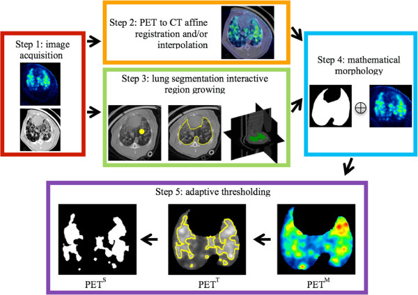
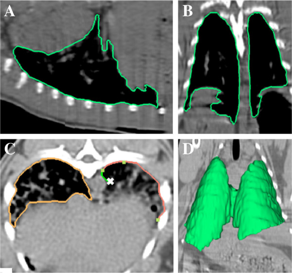
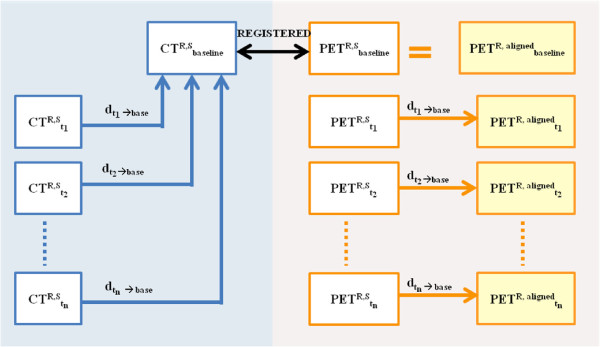
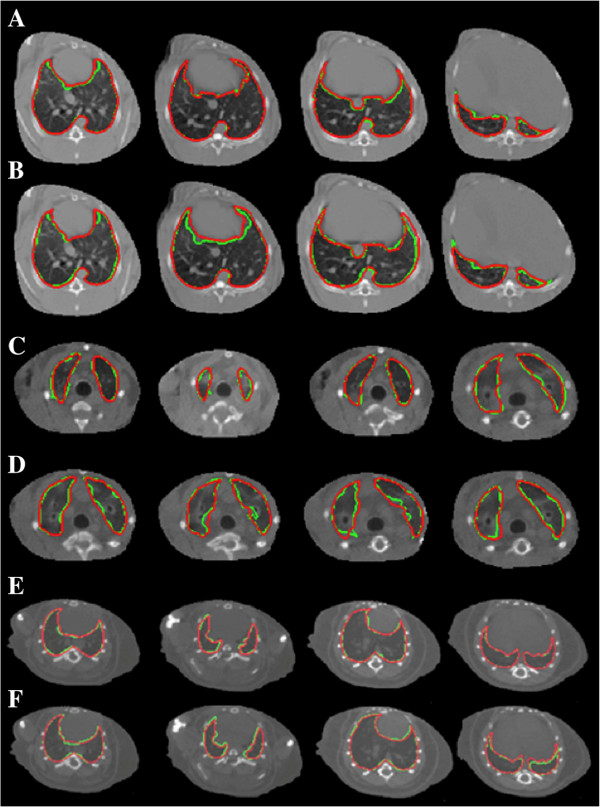
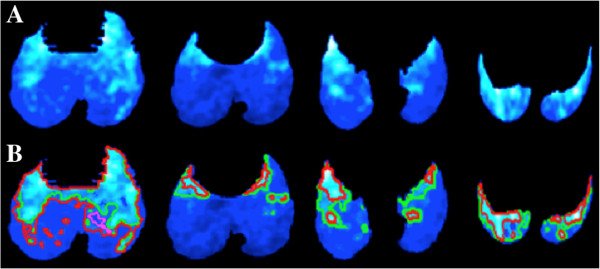
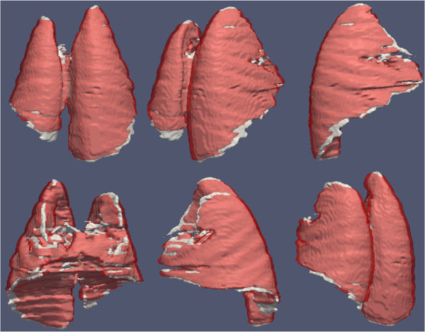
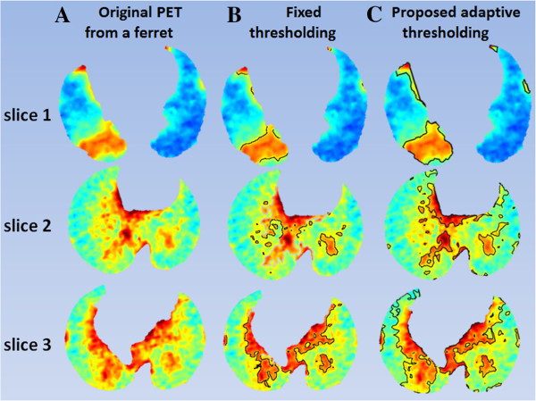
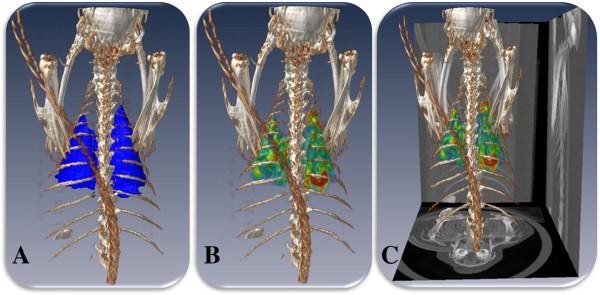
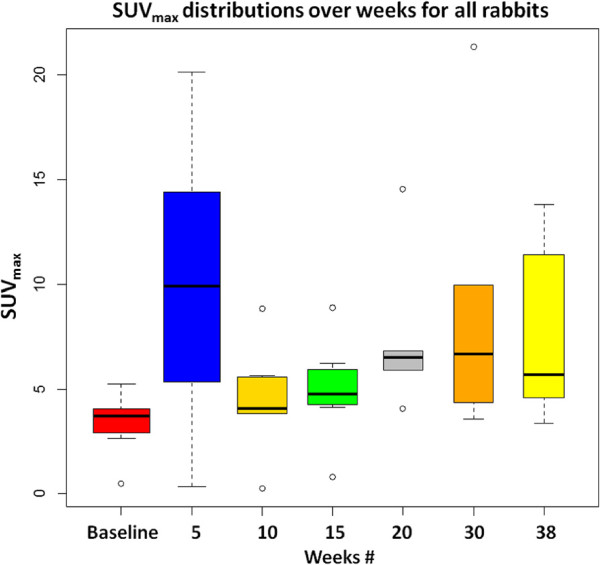
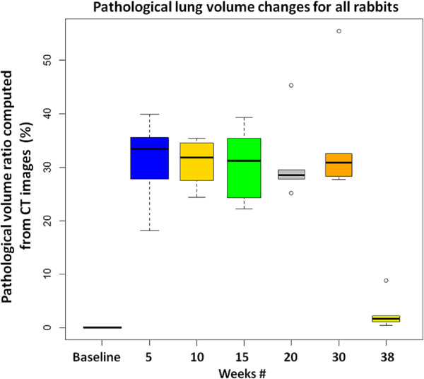
We tested the utility of our proposed methodology by using sequentially acquired CT and PET images of rabbit, ferret, and mouse models with respiratory infections of Mycobacterium tuberculosis (TB), H1N1 flu virus, and an aerosolized respiratory pathogen (necrotic TB) for a total of 92, 44, and 24 scans for the respective studies with half of the scans from CT and the other half from PET. Institutional Administrative Panel on Laboratory Animal Care approvals were obtained prior to conducting this research. First, the proposed computational framework registered PET and CT images to provide spatial correspondences between images. Second, the lungs from the CT scans were segmented using an interactive region growing (IRG) segmentation algorithm with mathematical morphology operations to avoid false positive (FP) uptake in PET images. Finally, we segmented significant radiotracer uptake from the PET images in lung regions determined from CT and computed metabolic volumes of the significant uptake. All segmentation processes were compared with expert radiologists' delineations (ground truths). Metabolic and gross volume of lesions were automatically computed with the segmentation processes using PET and CT images, and percentage changes in those volumes over time were calculated. (Continued on next page)(Continued from previous page) Standardized uptake value (SUV) analysis from PET images was conducted as a complementary quantitative metric for disease severity assessment. Thus, severity and extent of pulmonary lesions were examined through both PET and CT images using the aforementioned quantification metrics outputted from the proposed framework.
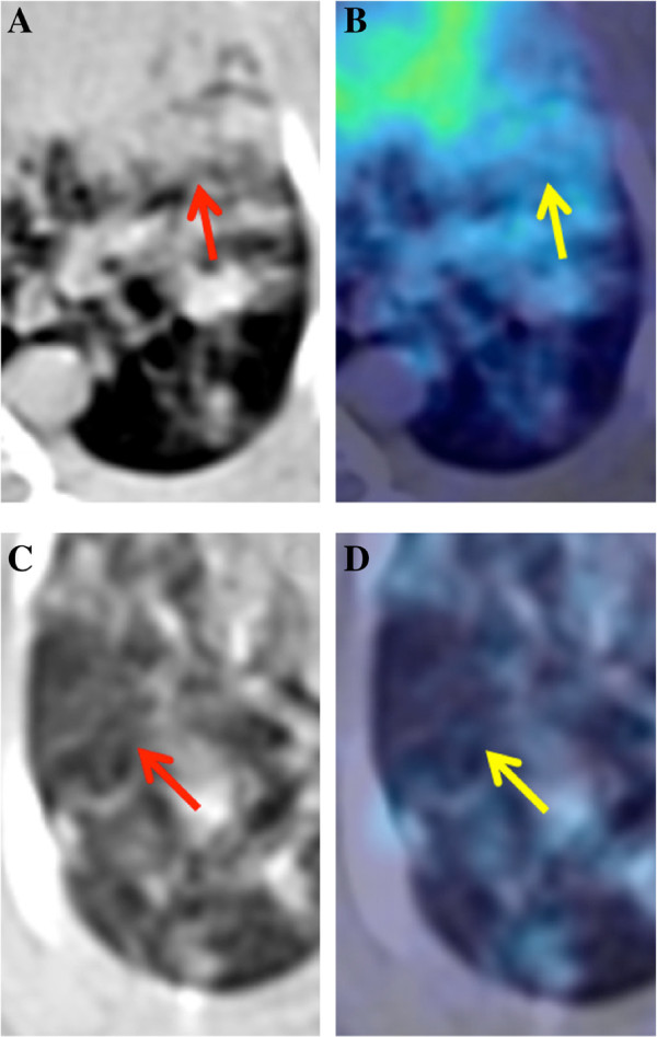
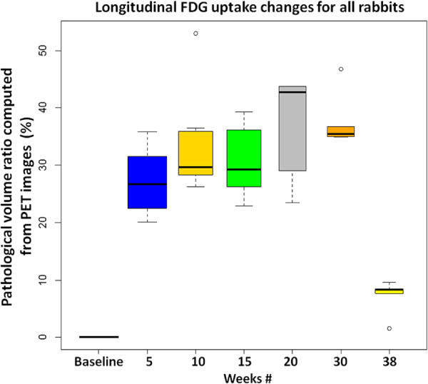
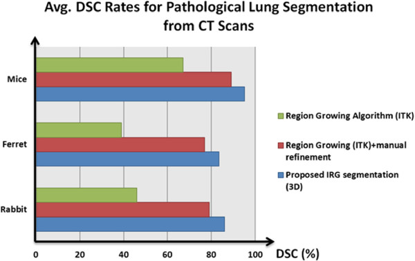
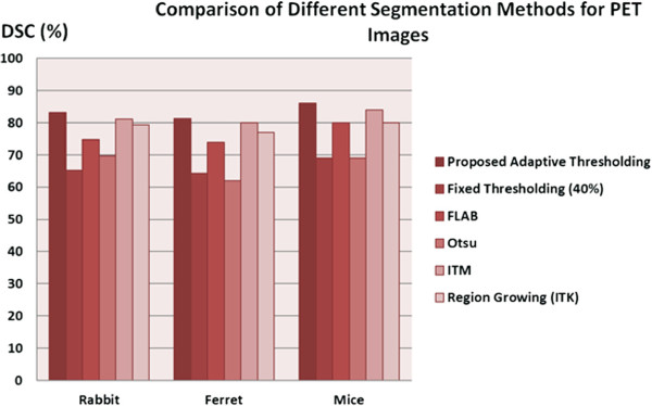
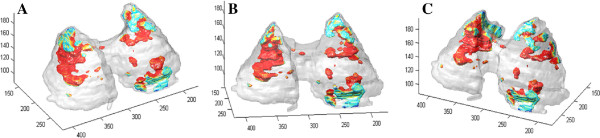
Each animal study was evaluated within the same subject class, and all steps of the proposed methodology were evaluated separately. We quantified the accuracy of the proposed algorithm with respect to the state-of-the-art segmentation algorithms. For evaluation of the segmentation results, dice similarity coefficient (DSC) as an overlap measure and Haussdorf distance as a shape dissimilarity measure were used. Significant correlations regarding the estimated lesion volumes were obtained both in CT and PET images with respect to the ground truths (R2=0.8922,p<0.01 and R2=0.8664,p<0.01, respectively). The segmentation accuracy (DSC (%)) was 93.4±4.5% for normal lung CT scans and 86.0±7.1% for pathological lung CT scans. Experiments showed excellent agreements (all above 85%) with expert evaluations for both structural and functional imaging modalities. Apart from quantitative analysis of each animal, we also qualitatively showed how metabolic volumes were changing over time by examining serial PET/CT scans. Evaluation of the registration processes was based on precisely defined anatomical landmark points by expert clinicians. An average of 2.66, 3.93, and 2.52 mm errors was found in rabbit, ferret, and mouse data (all within the resolution limits), respectively. Quantitative results obtained from the proposed methodology were visually related to the progress and severity of the pulmonary infections as verified by the participating radiologists. Moreover, we demonstrated that lesions due to the infections were metabolically active and appeared multi-focal in nature, and we observed similar patterns in the CT images as well. Consolidation and ground glass opacity were the main abnormal imaging patterns and consistently appeared in all CT images. We also found that the gross and metabolic lesion volume percentage follow the same trend as the SUV-based evaluation in the longitudinal analysis.
We explored the feasibility of using PET and CT imaging modalities in three distinct small animal models for two diverse pulmonary infections. We concluded from the clinical findings, derived from the proposed computational pipeline, that PET-CT imaging is an invaluable hybrid modality for tracking pulmonary infections longitudinally in small animals and has great potential to become routinely used in clinics. Our proposed methodology showed that automated computed-aided lesion detection and quantification of pulmonary infections in small animal models are efficient and accurate as compared to the clinical standard of manual and semi-automated approaches. Automated analysis of images in pre-clinical applications can increase the efficiency and quality of pre-clinical findings that ultimately inform downstream experimental design in human clinical studies; this innovation will allow researchers and clinicians to more effectively allocate study resources with respect to research demands without compromising accuracy.
传染病是全球第二大致死原因。为了更好地理解和治疗传染病,需要使用多模态成像技术对解剖结构和功能特征进行准确评估。对于非侵入性成像技术,如计算机断层扫描(CT)、磁共振成像(MRI)和正电子发射断层扫描(PET),已经有许多工程改进,显著提高了图像的分辨率和对比度,但仍然缺乏研究人员在准确量化解剖结构和功能生物过程的成像数据时可用的计算算法。由于这些工具的开发可能潜在地将基础研究转化为临床实践,因此本研究专注于开发一种定量和定性的图像分析平台,为小动物模型中的肺部感染提供计算放射学视角。具体而言,我们设计了 (a) 一个快速且稳健的自动化和半自动图像分析平台和量化工具,可方便准确地测量肺部病变的诊断测量值以及解剖结构的体积测量值,并纳入了 (b) 图像配准管道,用于我们提出的框架,以对系列扫描进行体积比较。这是一种用于小动物传染病模型的重要研究工具,可以帮助研究人员深入了解传染病。
我们通过使用具有结核分枝杆菌(TB)、H1N1 流感病毒和雾化呼吸道病原体(坏死性 TB)呼吸道感染的兔、雪貂和小鼠模型的连续获取的 CT 和 PET 图像来测试我们提出的方法的效用,每个研究的总扫描次数分别为 92、44 和 24 次,其中一半来自 CT,另一半来自 PET。在进行这项研究之前,获得了机构行政动物护理小组的批准。首先,拟议的计算框架对 PET 和 CT 图像进行配准,以提供图像之间的空间对应关系。其次,使用交互式区域生长(IRG)分割算法和数学形态学操作对 CT 扫描中的肺部进行分割,以避免 PET 图像中的假阳性(FP)摄取。最后,我们从 CT 确定的肺部区域中分割出来自 PET 图像的显著放射性摄取,并计算出显著摄取的代谢体积。所有分割过程都与专家放射科医生的描绘(地面真实)进行了比较。使用 PET 和 CT 图像的分割过程自动计算病变的代谢和总体积,计算出随时间变化的体积百分比变化。(续页)(续前页)从 PET 图像进行标准化摄取值(SUV)分析是作为疾病严重程度评估的补充定量指标。因此,通过从提出的框架输出的上述量化指标,使用 PET 和 CT 图像检查了肺部病变的严重程度和范围。
对每个动物研究在同一类别内进行评估,并分别评估了所提出方法的所有步骤。我们使用重叠度量的骰子相似系数(DSC)和形状差异度量的 Haussdorf 距离来量化算法的准确性。对于 CT 和 PET 图像,都获得了与地面真实值具有显著相关性的估计病变体积(R2=0.8922,p<0.01 和 R2=0.8664,p<0.01,分别)。正常肺部 CT 扫描的分割准确性(DSC(%))为 93.4±4.5%,而病理肺部 CT 扫描的分割准确性为 86.0±7.1%。实验结果与专家评估在结构和功能成像模态方面都具有出色的一致性(均高于 85%)。除了对每个动物的定量分析外,我们还通过检查连续的 PET/CT 扫描,定性地展示了代谢体积随时间的变化情况。配准过程的评估基于专家临床医生精确定义的解剖学地标点。在兔、雪貂和小鼠数据中,分别发现平均 2.66、3.93 和 2.52 毫米的误差(均在分辨率限制内)。从提出的方法获得的定量结果在与放射科医生一起验证的临床发现中与肺部感染的进展和严重程度相关,并且,我们证明了感染引起的病变具有代谢活性,并且呈现多灶性,并且在 CT 图像中也观察到了类似的模式。实变和磨玻璃影是主要的异常成像模式,并且在所有 CT 图像中都一致出现。我们还发现,在纵向分析中,总病变和代谢病变体积百分比与基于 SUV 的评估具有相同的趋势。
我们探索了使用两种不同的肺部感染小动物模型中的 PET 和 CT 成像方式的可行性。我们从提出的计算管道得出的临床发现得出结论,PET-CT 成像对于在小动物中进行纵向跟踪肺部感染是一种非常有价值的混合模态,并且具有成为临床常规使用的巨大潜力。与手动和半自动方法相比,我们提出的方法在小动物模型中的肺部感染的自动计算机辅助病变检测和量化方面显示出高效和准确。在临床前应用中进行图像的自动分析可以提高临床前研究的效率和质量,最终为人类临床研究中的下游实验设计提供信息,这一创新将使研究人员和临床医生能够更有效地分配研究资源,而不会牺牲准确性。