Cardiorenal Research Laboratory, Department of Vascular Advanced Medicine, Faculty of Medicine, University of Miyazaki, 5200 Kihara, Kiyotake, Miyazaki, 889-1692, Japan.
Division of Internal Medicine, Cardiovascular Medicine and Nephrology, Faculty of Medicine, University of Miyazaki, 5200 Kihara, Kiyotake, Miyazaki, 889-1692, Japan.
Arch Osteoporos. 2023 Oct 24;18(1):129. doi: 10.1007/s11657-023-01339-2.
The purpose of this study was to investigate the morphological characteristics of the aortic valve and identify factors associated with the progression of aortic valve stenosis (AS) in osteoporosis patients.
In this single-center prospective cohort study, we recruited 10 patients (mean age: 75 ± 7 years, 90% female) who were taking anti-resorptive medicines at the outpatient clinic of University of Miyazaki Hospital, Japan. Baseline assessments, including transthoracic echocardiogram, blood sampling, and dual energy X-ray absorptiometry, were performed. Follow-up assessments were conducted at 6, 12, 18, and 24 months.
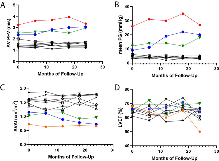
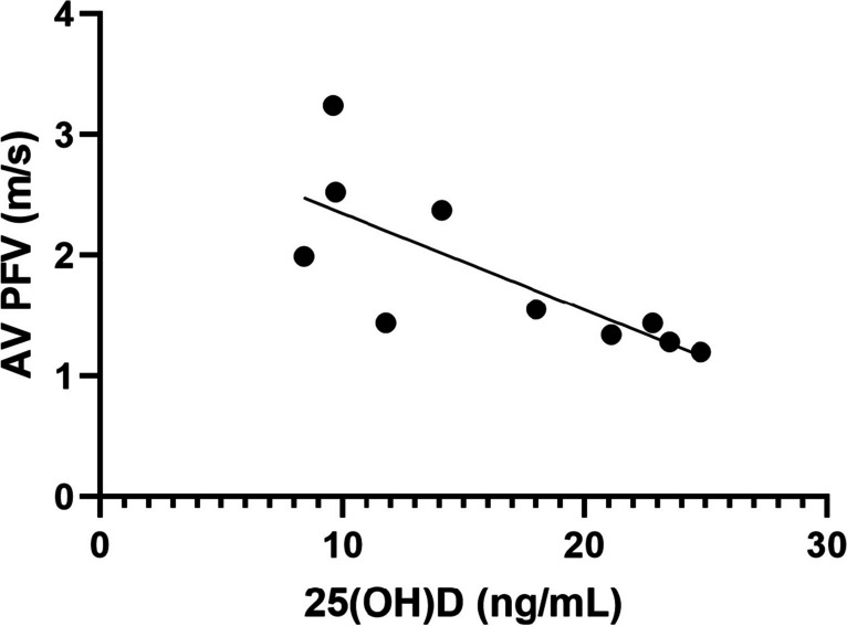
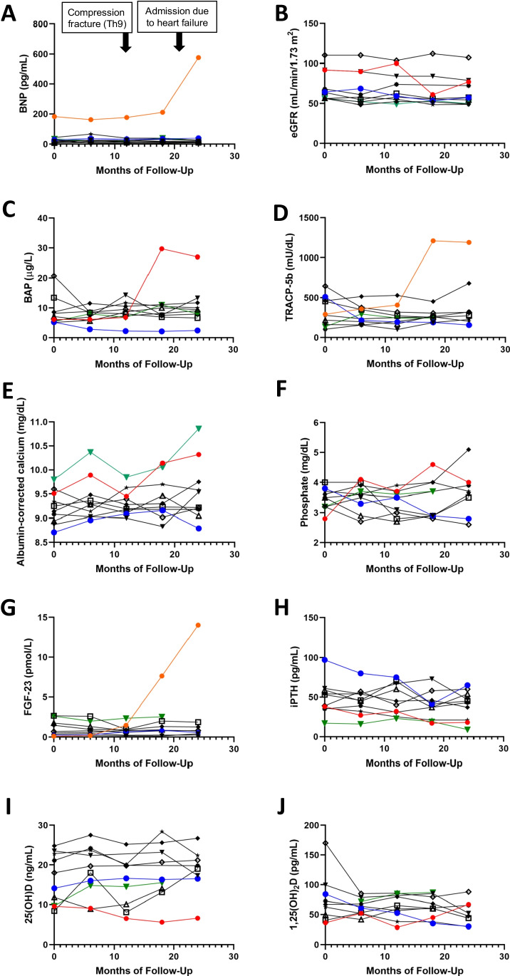
During the 2-year follow-up, three patients with aortic valve peak flow velocity (AV PFV) ≥2 m/s at baseline developed moderate AS, which is defined as AV PFV ≥3 m/s. However, seven patients with AV PFV <2 m/s did not exhibit any progression of AS. There were significant variations in terms of bone mineral density, T-score values, and biomarkers associated with bone turnover (i.e., bone alkaline phosphatase, tartrate-resistance acid phosphatase-5b) among the enrolled patients, but none of these factors were found to be associated with the progression of AS. All patients exhibited low vitamin D status, with a median level of 16.1 ng/mL (25 percentile, 9.7 ng/mL; 75 percentile, 23 ng/mL). The baseline levels of AV PFV values were negatively correlated with 25-hydroxyvitamin D levels, determined by univariate linear regression analysis (beta coefficient = -0.756, 95% confidence interval, -0.136 ̶ -0.023, p = 0.011).
Our data suggest that low vitamin D status might be a potential risk factor for the progression of AS in osteoporosis patients undergoing treatment with anti-resorptive medicines. Elderly patients with osteoporosis patients exhibited a subset of aortic valve stenosis. Our data suggest that the baseline aortic valve peak flow velocity predicts the progression of aortic valve stenosis, and there might be an association between the progression and the co-existing low vitamin D status in these patients.
本研究旨在探讨主动脉瓣的形态学特征,并确定与骨质疏松症患者主动脉瓣狭窄(AS)进展相关的因素。
本单中心前瞻性队列研究纳入了在日本宫崎大学医院门诊接受抗吸收药物治疗的 10 名患者(平均年龄:75 ± 7 岁,90%为女性)。进行了基线评估,包括经胸超声心动图、血液采样和双能 X 线吸收法。在 6、12、18 和 24 个月时进行随访评估。
在 2 年的随访期间,基线时主动脉瓣峰值流速(AV PFV)≥2 m/s 的 3 名患者发展为中度 AS,定义为 AV PFV≥3 m/s。然而,7 名 AV PFV<2 m/s 的患者没有出现任何 AS 进展。入组患者的骨密度、T 评分值和与骨转换相关的生物标志物(即骨碱性磷酸酶、抗酒石酸酸性磷酸酶-5b)存在显著差异,但这些因素均与 AS 进展无关。所有患者均表现出低维生素 D 状态,中位水平为 16.1 ng/mL(25%位数,9.7 ng/mL;75%位数,23 ng/mL)。通过单变量线性回归分析发现,AV PFV 值的基线水平与 25-羟维生素 D 水平呈负相关(β系数=-0.756,95%置信区间,-0.136 ̶ -0.023,p=0.011)。
我们的数据表明,低维生素 D 状态可能是骨质疏松症患者接受抗吸收药物治疗时 AS 进展的潜在危险因素。接受抗吸收药物治疗的骨质疏松症患者存在亚组主动脉瓣狭窄。我们的数据表明,基线 AV PFV 预测 AS 的进展,并且这些患者的进展可能与共存的低维生素 D 状态有关。