Bureau Flavien, Robin Justine, Le Ber Arthur, Lambert William, Fink Mathias, Aubry Alexandre
Institut Langevin, ESPCI Paris, PSL University, CNRS, 75005, Paris, France.
Physics for Medicine, ESPCI Paris, PSL University, INSERM, CNRS, Paris, France.
Nat Commun. 2023 Oct 25;14(1):6793. doi: 10.1038/s41467-023-42338-8.
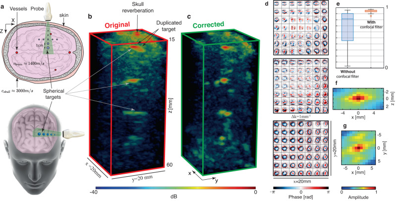
Matrix imaging paves the way towards a next revolution in wave physics. Based on the response matrix recorded between a set of sensors, it enables an optimized compensation of aberration phenomena and multiple scattering events that usually drastically hinder the focusing process in heterogeneous media. Although it gave rise to spectacular results in optical microscopy or seismic imaging, the success of matrix imaging has been so far relatively limited with ultrasonic waves because wave control is generally only performed with a linear array of transducers. In this paper, we extend ultrasound matrix imaging to a 3D geometry. Switching from a 1D to a 2D probe enables a much sharper estimation of the transmission matrix that links each transducer and each medium voxel. Here, we first present an experimental proof of concept on a tissue-mimicking phantom through ex-vivo tissues and then, show the potential of 3D matrix imaging for transcranial applications.
矩阵成像为波动物理学的下一次革命铺平了道路。基于一组传感器之间记录的响应矩阵,它能够对像差现象和多次散射事件进行优化补偿,而这些现象通常会严重阻碍异质介质中的聚焦过程。尽管它在光学显微镜或地震成像中取得了惊人的成果,但到目前为止,矩阵成像在超声波方面的成功相对有限,因为波控制通常仅通过线性换能器阵列来执行。在本文中,我们将超声矩阵成像扩展到三维几何结构。从一维探头切换到二维探头能够更精确地估计连接每个换能器和每个介质体素的传输矩阵。在这里,我们首先通过离体组织在组织模拟体模上展示了概念验证实验,然后展示了三维矩阵成像在经颅应用中的潜力。