Spelman L H, Thompson N L, Fausto N, Miller K R
Am J Pathol. 1986 Nov;125(2):379-92.
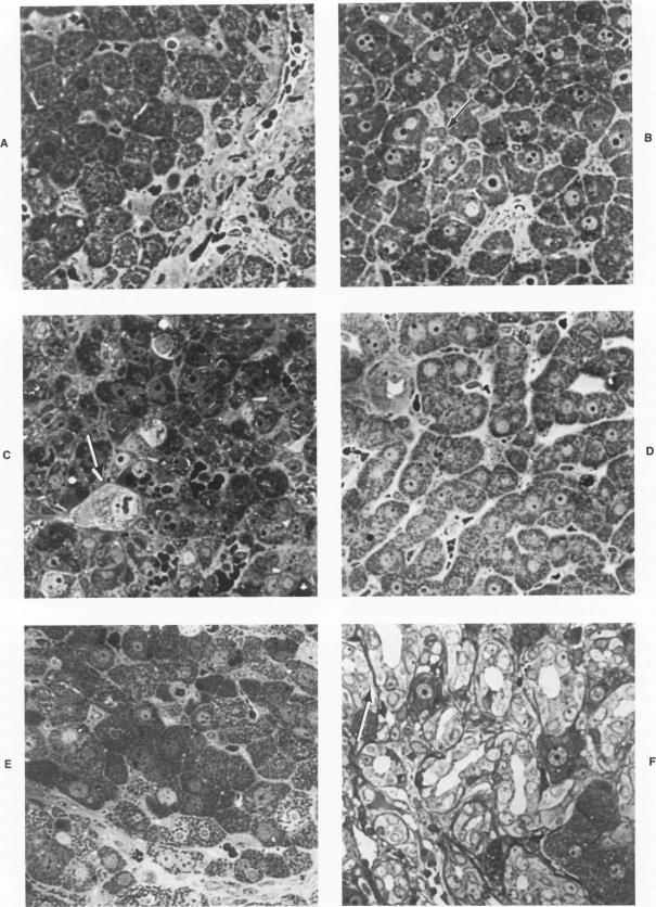
The authors have investigated early changes in liver cell gap and tight junctions that occur when rats are fed a carcinogenic diet. Animals were fed a choline-deficient diet that contained 0.1% ethionine (CDE) for periods up to 6 weeks. Short-term feeding of this diet results in the rapid proliferation of so-called "oval cells" within the liver, which is reversible upon returning the rats to a normal diet. Livers from animals fed the diet were removed at various times during feeding and during recovery from the diet and were analyzed by light and electron microscopy. The freeze-fracture technique was used to produce extended views of the internal structure of liver cell membranes at each stage under study. The characteristic junctional complex surrounding canalicular regions in normal liver disappears after only 2 weeks of the CDE regimen. Gap junctions were not found after 4 weeks of the diet, and tight junctions became increasingly disorganized. Tight junction elements were observed, however, between hepatocytes and oval cells, which indicated that these two cell types do interact directly. Changes occur in the structural complexity of tight junction elements between hepatocytes and between hepatocytes and oval cells. Recovery from the CDE diet results in a rapid increase in junctional complexity, and the large gap junction plaques characteristic of normal liver are visible within 2 weeks after cessation of the CDE regimen. These and other observations demonstrate that reversible alterations in hepatocyte gap and tight junctions occur as a result of administration of a diet that induces oval cell proliferation. The relationship of these changes to those that have been reported during other processes of cell proliferation are discussed.
作者研究了给大鼠喂食致癌饮食时肝细胞间隙连接和紧密连接的早期变化。动物被喂食含有0.1%乙硫氨酸的胆碱缺乏饮食(CDE),持续时间长达6周。短期喂食这种饮食会导致肝脏内所谓“卵圆细胞”的快速增殖,当大鼠恢复正常饮食后这种增殖是可逆的。在喂食期间以及从饮食中恢复的不同时间,取出喂食该饮食的动物的肝脏,通过光镜和电镜进行分析。在研究的每个阶段,使用冷冻断裂技术来呈现肝细胞细胞膜内部结构的扩展视图。正常肝脏中围绕胆小管区域的特征性连接复合体在CDE方案仅2周后就消失了。饮食4周后未发现间隙连接,紧密连接变得越来越紊乱。然而,在肝细胞和卵圆细胞之间观察到紧密连接成分,这表明这两种细胞类型确实直接相互作用。肝细胞之间以及肝细胞与卵圆细胞之间紧密连接成分的结构复杂性发生了变化。从CDE饮食中恢复导致连接复杂性迅速增加,在停止CDE方案2周内可见正常肝脏特有的大间隙连接斑块。这些以及其他观察结果表明,由于给予诱导卵圆细胞增殖的饮食,肝细胞间隙连接和紧密连接发生了可逆变化。讨论了这些变化与其他细胞增殖过程中所报道变化的关系。