Huang Coco X, Siwan Elisha, Fox Sarah L, Longfield Matilda, Twigg Stephen M, Min Danqing
Greg Brown Diabetes and Endocrine Research Laboratory, Sydney Medical School (Central), Faculty of Medicine and Health, Charles Perkins Centre, The University of Sydney, Sydney, NSW, Australia.
Department of Endocrinology, Royal Prince Alfred Hospital, Sydney, NSW, Australia.
Lab Anim Res. 2023 Oct 27;39(1):25. doi: 10.1186/s42826-023-00176-1.
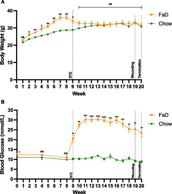
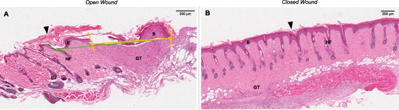
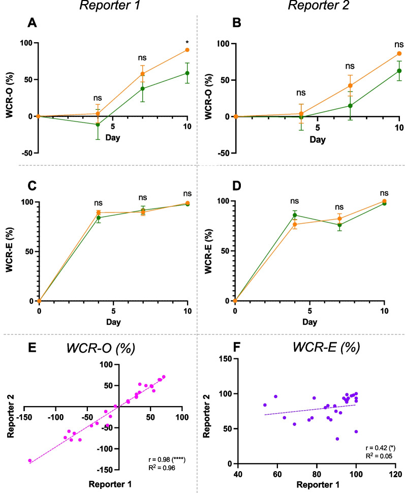
Chronic skin wounds are a common complication of many diseases such as diabetes. Various traditional methods for assessing skin wound closure are used in animal studies, including wound tracing, calliper measurements and histological analysis. However, these methods have poorly defined wound closure or practical limitations. Digital image analysis of wounds is an increasingly popular, accessible alternative, but it is unclear whether digital assessment is consistent with traditional methods. This study aimed to optimise and compare digital wound closure assessment with traditional methods, using a diabetic mouse model. Diabetes was induced in male C57BL/6J mice by high-fat diet feeding combined with low dose (65 mg/kg of body weight) streptozotocin injections. Mice fed normal chow were included as controls. After 18 weeks, four circular full-thickness dorsal skin wounds of 4 mm diameter were created per mouse. The wounds were photographed and measured by callipers. Wound closure rate (WCR) was digitally assessed by two reporters using two methods: wound outline (WCR-O) and re-epithelialisation (WCR-E). Wounded skin tissues were collected at 10-days post-wounding and wound width was measured from haematoxylin and eosin-stained skin tissue.
Between reporters, WCR-O was more consistent than WCR-E, and WCR-O correlated with calliper measurements. Histological analysis supported digital assessments, especially WCR-E, when wounds were histologically closed.
WCR-O could replace calliper measurements to measure skin wound closure, but WCR-E assessment requires further refinement. Small animal studies of skin wound healing can greatly benefit from standardised definitions of wound closure and more consistent digital assessment protocols.
慢性皮肤伤口是许多疾病(如糖尿病)的常见并发症。在动物研究中使用了各种评估皮肤伤口愈合的传统方法,包括伤口追踪、卡尺测量和组织学分析。然而,这些方法对伤口愈合的定义不明确或存在实际局限性。伤口的数字图像分析是一种越来越受欢迎且易于使用的替代方法,但尚不清楚数字评估是否与传统方法一致。本研究旨在使用糖尿病小鼠模型优化数字伤口愈合评估并与传统方法进行比较。通过高脂饮食喂养结合低剂量(65mg/kg体重)链脲佐菌素注射诱导雄性C57BL/6J小鼠患糖尿病。喂食正常饲料的小鼠作为对照。18周后,每只小鼠在背部制造4个直径为4mm的圆形全层皮肤伤口。对伤口进行拍照并用卡尺测量。由两名记录员使用两种方法对伤口愈合率(WCR)进行数字评估:伤口轮廓(WCR-O)和再上皮化(WCR-E)。在受伤后10天收集受伤的皮肤组织,并从苏木精和伊红染色的皮肤组织中测量伤口宽度。
在记录员之间,WCR-O比WCR-E更一致,并且WCR-O与卡尺测量结果相关。组织学分析支持数字评估,尤其是当伤口在组织学上愈合时的WCR-E。
WCR-O可以替代卡尺测量来测量皮肤伤口愈合,但WCR-E评估需要进一步完善。皮肤伤口愈合的小动物研究可以从标准化的伤口愈合定义和更一致的数字评估方案中大大受益。