• 文献检索
  • 文档翻译
  • 深度研究
  • 学术资讯
  • Suppr Zotero 插件Zotero 插件
  • 邀请有礼
  • 套餐&价格
  • 历史记录
应用&插件
Suppr Zotero 插件Zotero 插件浏览器插件Mac 客户端Windows 客户端微信小程序
定价
高级版会员购买积分包购买API积分包
服务
文献检索文档翻译深度研究API 文档MCP 服务
关于我们
关于 Suppr公司介绍联系我们用户协议隐私条款
关注我们

Suppr 超能文献

核心技术专利:CN118964589B侵权必究
粤ICP备2023148730 号-1Suppr @ 2026

文献检索

告别复杂PubMed语法,用中文像聊天一样搜索,搜遍4000万医学文献。AI智能推荐,让科研检索更轻松。

立即免费搜索

文件翻译

保留排版,准确专业,支持PDF/Word/PPT等文件格式,支持 12+语言互译。

免费翻译文档

深度研究

AI帮你快速写综述,25分钟生成高质量综述,智能提取关键信息,辅助科研写作。

立即免费体验

革兰氏阴性菌细胞外囊泡定量方法的比较。

Comparison of Methods for Quantifying Extracellular Vesicles of Gram-Negative Bacteria.

机构信息

Microbiology and Cell Science Department, Institute of Food and Agricultural Sciences, University of Florida, Gainesville, FL 32611, USA.

出版信息

Int J Mol Sci. 2023 Oct 11;24(20):15096. doi: 10.3390/ijms242015096.

DOI:10.3390/ijms242015096
PMID:37894776
原文链接:https://pmc.ncbi.nlm.nih.gov/articles/PMC10606555/
Abstract

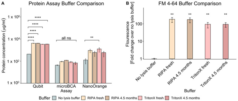
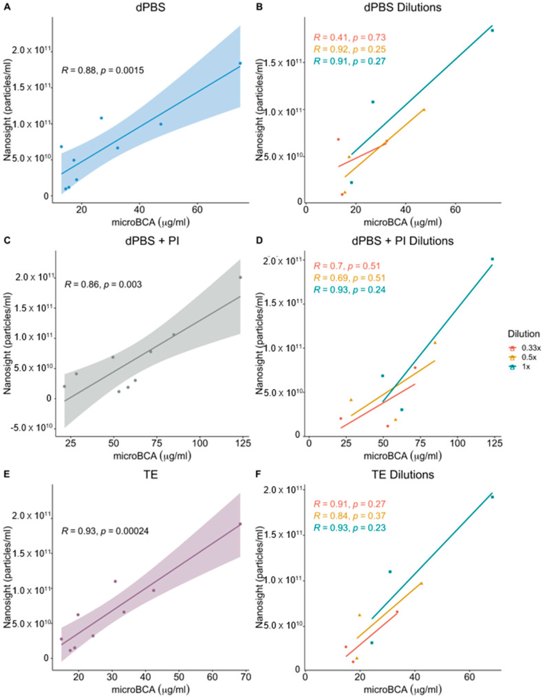
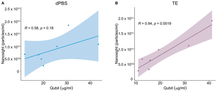
There are a variety of methods employed by laboratories for quantifying extracellular vesicles isolated from bacteria. As a result, the ability to compare results across published studies can lead to questions regarding the suitability of methods and buffers for accurately quantifying these vesicles. Within the literature, there are several common methods for vesicle quantification. These include lipid quantification using the lipophilic dye FM 4-64, protein quantification using microBCA, Qubit, and NanoOrange assays, or direct vesicle enumeration using nanoparticle tracking analysis. In addition, various diluents and lysis buffers are also used to resuspend and treat vesicles. In this study, we directly compared the quantification of a bacterial outer membrane vesicle using several commonly used methods. We also tested the impact of different buffers, buffer age, lysis method, and vesicle diluent on vesicle quantification. The results showed that buffer age had no significant effect on vesicle quantification, but the lysis method impacted the reliability of measurements using Qubit and NanoOrange. The microBCA assay displayed the least variability in protein concentration values and was the most consistent, regardless of the buffer or diluent used. MicroBCA also demonstrated the strongest correlation to the NTA-determined particle number across a range of vesicle concentrations. Overall, these results indicate that with appropriate diluent and buffer choice, microBCA vs. NTA standard curves could be generated and the microBCA assay used to estimate the particle number when NTA instrumentation is not readily available.

摘要

实验室采用了多种方法来定量从细菌中分离出的细胞外囊泡。因此,比较已发表研究结果的能力会引发对方法和缓冲液是否适合准确定量这些囊泡的质疑。在文献中,有几种常见的囊泡定量方法。这些方法包括使用亲脂性染料 FM 4-64 进行脂质定量、使用微 BCA、Qubit 和 NanoOrange 测定法进行蛋白质定量,或使用纳米颗粒跟踪分析直接对囊泡进行计数。此外,还使用各种稀释剂和裂解缓冲液来重悬和处理囊泡。在这项研究中,我们直接比较了几种常用方法对细菌外膜囊泡的定量。我们还测试了不同缓冲液、缓冲液老化、裂解方法和囊泡稀释剂对囊泡定量的影响。结果表明,缓冲液老化对囊泡定量没有显著影响,但裂解方法会影响 Qubit 和 NanoOrange 测量的可靠性。微 BCA 测定法在蛋白质浓度值的变化方面表现出最小的变异性,并且无论使用哪种缓冲液或稀释剂都最一致。微 BCA 与 NTA 确定的粒子数在一系列囊泡浓度下也具有最强的相关性。总体而言,这些结果表明,通过适当的稀释剂和缓冲液选择,可以生成微 BCA 与 NTA 标准曲线,并在没有 NTA 仪器的情况下使用微 BCA 测定法来估计粒子数。

https://cdn.ncbi.nlm.nih.gov/pmc/blobs/5d9f/10606555/8663960c36b2/ijms-24-15096-g004.jpg
https://cdn.ncbi.nlm.nih.gov/pmc/blobs/5d9f/10606555/ccb5e10052c5/ijms-24-15096-g001.jpg
https://cdn.ncbi.nlm.nih.gov/pmc/blobs/5d9f/10606555/1ca8ede16a3b/ijms-24-15096-g002.jpg
https://cdn.ncbi.nlm.nih.gov/pmc/blobs/5d9f/10606555/db07aed86cd5/ijms-24-15096-g003.jpg
https://cdn.ncbi.nlm.nih.gov/pmc/blobs/5d9f/10606555/8663960c36b2/ijms-24-15096-g004.jpg

相似文献

1
Comparison of Methods for Quantifying Extracellular Vesicles of Gram-Negative Bacteria.革兰氏阴性菌细胞外囊泡定量方法的比较。
Int J Mol Sci. 2023 Oct 11;24(20):15096. doi: 10.3390/ijms242015096.
2
Considerations for the Analysis of Bacterial Membrane Vesicles: Methods of Vesicle Production and Quantification Can Influence Biological and Experimental Outcomes.考虑细菌膜泡的分析:囊泡生产和定量的方法会影响生物学和实验结果。
Microbiol Spectr. 2021 Dec 22;9(3):e0127321. doi: 10.1128/Spectrum.01273-21.
3
Improving Reproducibility to Meet Minimal Information for Studies of Extracellular Vesicles 2018 Guidelines in Nanoparticle Tracking Analysis.提高重复性以满足细胞外囊泡研究的最小信息 2018 年纳米颗粒跟踪分析指南。
J Vis Exp. 2021 Nov 17(177). doi: 10.3791/63059.
4
Enumeration of extracellular vesicles by a new improved flow cytometric method is comparable to fluorescence mode nanoparticle tracking analysis.通过一种新的改进的流式细胞术方法对细胞外囊泡进行计数,与荧光模式纳米颗粒跟踪分析相当。
Nanomedicine. 2016 May;12(4):977-986. doi: 10.1016/j.nano.2015.12.370. Epub 2016 Jan 6.
5
Differential fluorescence nanoparticle tracking analysis for enumeration of the extracellular vesicle content in mixed particulate solutions.基于差分荧光纳米颗粒跟踪分析的混合颗粒体系中外泌体含量的检测。
Methods. 2020 May 1;177:67-73. doi: 10.1016/j.ymeth.2020.02.006. Epub 2020 Feb 17.
6
Stress-Inducible Protein 1 (STI1): Extracellular Vesicle Analysis and Quantification.应激诱导蛋白1(STI1):细胞外囊泡分析与定量
Methods Mol Biol. 2016;1459:161-74. doi: 10.1007/978-1-4939-3804-9_11.
7
FRET-Based Assay for the Quantification of Extracellular Vesicles and Other Vesicles of Complex Composition.基于荧光共振能量转移的用于定量细胞外囊泡和其他复杂组成囊泡的方法。
Anal Chem. 2020 Dec 1;92(23):15336-15343. doi: 10.1021/acs.analchem.0c02271. Epub 2020 Nov 12.
8
Systematic Evaluation of PKH Labelling on Extracellular Vesicle Size by Nanoparticle Tracking Analysis.通过纳米颗粒跟踪分析对细胞外囊泡大小进行 PKH 标记的系统评价。
Sci Rep. 2020 Jun 12;10(1):9533. doi: 10.1038/s41598-020-66434-7.
9
Nanoparticle Tracking Analysis for the Quantification and Size Determination of Extracellular Vesicles.纳米颗粒跟踪分析用于定量和大小测定细胞外囊泡。
J Vis Exp. 2021 Mar 28(169). doi: 10.3791/62447.
10
Gram-negative and Gram-positive bacterial extracellular vesicles.革兰氏阴性菌和革兰氏阳性菌的细胞外囊泡。
Semin Cell Dev Biol. 2015 Apr;40:97-104. doi: 10.1016/j.semcdb.2015.02.006. Epub 2015 Feb 19.

引用本文的文献

1
Therapeutic potential of mesenchymal stem cell-derived extracellular vesicles: A focus on inflammatory bowel disease.间充质干细胞衍生的细胞外囊泡的治疗潜力:以炎症性肠病为重点。
Clin Transl Med. 2024 Nov;14(11):e70075. doi: 10.1002/ctm2.70075.
2
Bacterial outer membrane vesicles in the fight against cancer.用于抗癌的细菌外膜囊泡
Chin Med J (Engl). 2024 Aug 9;137(18):2169-81. doi: 10.1097/CM9.0000000000003234.
3
Introduction to the Special Issue Dedicated to Extracellular Vesicles and Nanoparticles, Part 1.专刊介绍 第一部分:细胞外囊泡和纳米粒子

本文引用的文献

1
Comparison of interferometric light microscopy with nanoparticle tracking analysis for the study of extracellular vesicles and bacteriophages.用于细胞外囊泡和噬菌体研究的干涉光显微镜与纳米颗粒跟踪分析的比较。
J Extracell Biol. 2023 Feb 23;2(2):e75. doi: 10.1002/jex2.75. eCollection 2023 Feb.
2
Role of Bacterial Extracellular Vesicles in Manipulating Infection.细菌细胞外囊泡在操纵感染中的作用。
Infect Immun. 2023 May 16;91(5):e0043922. doi: 10.1128/iai.00439-22. Epub 2023 Apr 25.
3
Murine Norovirus Interaction with Enterobacter cloacae Leads to Changes in Membrane Stability and Packaging of Lipid and Metabolite Vesicle Content.
Int J Mol Sci. 2024 Jul 17;25(14):7805. doi: 10.3390/ijms25147805.
鼠诺如病毒与阴沟肠杆菌的相互作用导致膜稳定性以及脂质和代谢物囊泡内容物包装的变化。
Microbiol Spectr. 2023 Mar 21;11(2):e0469122. doi: 10.1128/spectrum.04691-22.
4
Assessment of Small Cellular Particles from Four Different Natural Sources and Liposomes by Interferometric Light Microscopy.利用干涉式荧光显微镜评估四种不同天然来源的小细胞颗粒和脂质体。
Int J Mol Sci. 2022 Dec 13;23(24):15801. doi: 10.3390/ijms232415801.
5
Bacterial extracellular vesicles control murine norovirus infection through modulation of antiviral immune responses.细菌细胞外囊泡通过调节抗病毒免疫应答来控制鼠诺如病毒感染。
Front Immunol. 2022 Aug 4;13:909949. doi: 10.3389/fimmu.2022.909949. eCollection 2022.
6
Isolation and characterization of extracellular vesicles and future directions in diagnosis and therapy.细胞外囊泡的分离与鉴定及其在诊断与治疗中的未来方向。
Wiley Interdiscip Rev Nanomed Nanobiotechnol. 2023 Jan;15(1):e1835. doi: 10.1002/wnan.1835. Epub 2022 Jul 27.
7
Proteomic Network of Antibiotic-Induced Outer Membrane Vesicles Released by Extensively Drug-Resistant Elizabethkingia anophelis.广泛耐药嗜水气单胞菌诱导的外膜囊泡的蛋白质组学网络。
Microbiol Spectr. 2022 Aug 31;10(4):e0026222. doi: 10.1128/spectrum.00262-22. Epub 2022 Jul 19.
8
Overexpression of the Bam Complex Improves the Production of MOMP in the Outer Membrane. Bam 复合物过表达提高外膜中 MOMP 的产量。
Int J Mol Sci. 2022 Jul 2;23(13):7393. doi: 10.3390/ijms23137393.
9
Lipidomics Analysis of Outer Membrane Vesicles and Elucidation of the Inositol Phosphoceramide Biosynthetic Pathway in Bacteroides thetaiotaomicron.外膜囊泡的脂质组学分析及拟杆菌属中肌醇磷酸神经酰胺生物合成途径的阐明。
Microbiol Spectr. 2022 Feb 23;10(1):e0063421. doi: 10.1128/spectrum.00634-21. Epub 2022 Jan 26.
10
An Intranasal OMV-Based Vaccine Induces High Mucosal and Systemic Protecting Immunity Against a SARS-CoV-2 Infection.一种基于鼻腔内的 OMV 疫苗可诱导针对 SARS-CoV-2 感染的高黏膜和全身保护免疫。
Front Immunol. 2021 Dec 17;12:781280. doi: 10.3389/fimmu.2021.781280. eCollection 2021.