Weill Institute for Neurosciences, Department of Neurology, University of California, San Francisco, San Francisco, CA, USA.
Neuroimaging Program, Department of Neurology, Cedars-Sinai Medical Center, Los Angeles, CA, USA.
Mult Scler. 2023 Dec;29(14):1736-1747. doi: 10.1177/13524585231204414. Epub 2023 Oct 28.
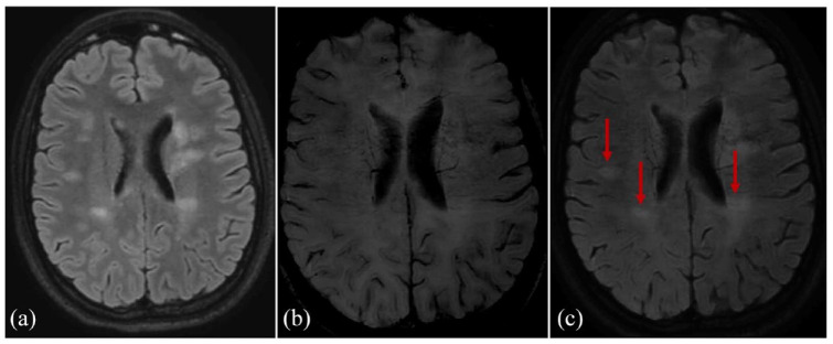
Myelin oligodendrocyte glycoprotein (MOG) antibody-associated disease (MOGAD) and pediatric-onset multiple sclerosis (POMS) share clinical and magnetic resonance imaging (MRI) features but differ in prognosis and management. Early POMS diagnosis is essential to avoid disability accumulation. Central vein sign (CVS), paramagnetic rim lesions (PRLs), and central core lesions (CCLs) are susceptibility-based imaging (SbI)-related signs understudied in pediatric populations that may help discerning POMS from MOGAD.
T2-FLAIR and SbI (three-dimensional echoplanar imaging (3D-EPI)/susceptibility-weighted imaging (SWI) or similar) were acquired on 1.5T/3T scanners. Two readers assessed CVS-positive rate (%CVS+), and their average score was used to build a receiver operator curve (ROC) assessing the ability to discriminate disease type. PRLs and CCLs were identified using a consensual approach.
The %CVS+ distinguished 26 POMS cases (mean age 13.7 years, 63% females, median EDSS 1.5) from 14 MOGAD cases (10.8 years, 35% females, EDSS 1.0) with ROC = 1, < 0.0001, (cutoff 41%). PRLs were only detectable in POMS participants (mean 2.1±2.3, range 1-10), discriminating the two conditions with a sensitivity of 69% and a specificity of 100%. CCLs were more sensitive (81%) but less specific (71.43%).
The %CVS+ and PRLs are highly specific markers of POMS. After proper validation on larger multicenter cohorts, consideration should be given to including such imaging markers for diagnosing POMS at disease onset.
髓鞘少突胶质细胞糖蛋白(MOG)抗体相关疾病(MOGAD)和儿童发病多发性硬化症(POMS)具有相似的临床和磁共振成像(MRI)特征,但在预后和治疗方面有所不同。早期 POMS 的诊断对于避免残疾累积至关重要。中央静脉征(CVS)、顺磁性边缘病变(PRLs)和中央核心病变(CCLs)是在儿科人群中研究较少的基于敏感性的成像(SbI)相关征象,这些征象可能有助于区分 POMS 和 MOGAD。
在 1.5T/3T 扫描仪上采集 T2-FLAIR 和 SbI(三维平面回波成像(3D-EPI)/磁敏感加权成像(SWI)或类似物)。两名读者评估了 CVS 阳性率(%CVS+),并使用他们的平均评分构建了一条评估区分疾病类型能力的接收者操作特征曲线(ROC)。使用共识方法确定了 PRLs 和 CCLs。
%CVS+区分了 26 例 POMS 病例(平均年龄 13.7 岁,63%为女性,EDSS 中位数为 1.5)和 14 例 MOGAD 病例(平均年龄 10.8 岁,35%为女性,EDSS 为 1.0),ROC = 1,<0.0001(截断值为 41%)。PRLs 仅在 POMS 参与者中可检测到(平均 2.1±2.3,范围 1-10),其敏感性为 69%,特异性为 100%。CCLs 的敏感性更高(81%),但特异性较低(71.43%)。
%CVS+和 PRLs 是 POMS 的高度特异性标志物。在更大的多中心队列上进行适当验证后,应考虑将此类成像标志物纳入 POMS 的发病初始诊断。