• 文献检索
  • 文档翻译
  • 深度研究
  • 学术资讯
  • Suppr Zotero 插件Zotero 插件
  • 邀请有礼
  • 套餐&价格
  • 历史记录
应用&插件
Suppr Zotero 插件Zotero 插件浏览器插件Mac 客户端Windows 客户端微信小程序
定价
高级版会员购买积分包购买API积分包
服务
文献检索文档翻译深度研究API 文档MCP 服务
关于我们
关于 Suppr公司介绍联系我们用户协议隐私条款
关注我们

Suppr 超能文献

核心技术专利:CN118964589B侵权必究
粤ICP备2023148730 号-1Suppr @ 2026

文献检索

告别复杂PubMed语法,用中文像聊天一样搜索,搜遍4000万医学文献。AI智能推荐,让科研检索更轻松。

立即免费搜索

文件翻译

保留排版,准确专业,支持PDF/Word/PPT等文件格式,支持 12+语言互译。

免费翻译文档

深度研究

AI帮你快速写综述,25分钟生成高质量综述,智能提取关键信息,辅助科研写作。

立即免费体验

细菌视紫红质M412中间体的电子衍射分析。

Electron diffraction analysis of the M412 intermediate of bacteriorhodopsin.

作者信息

Glaeser R M, Baldwin J, Ceska T A, Henderson R

出版信息

Biophys J. 1986 Nov;50(5):913-20. doi: 10.1016/S0006-3495(86)83532-9.

DOI:10.1016/S0006-3495(86)83532-9
PMID:3790694
原文链接:https://pmc.ncbi.nlm.nih.gov/articles/PMC1329816/
Abstract

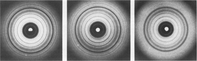
High resolution electron diffraction data have been recorded for glucose-embedded purple membrane specimens in which bacteriorhodopsin (bR) has been trapped by cooling slowly to below--100 degrees C under continuous illumination. Thin films (OD approximately 0.7) of glucose-embedded membranes, prepared as a control, showed virtually 100% conversion to the M state, and stacks of such thin film specimens gave very similar x-ray diffraction patterns in the bR568 and the M412 state in most experiments. To be certain that any measured differences in diffraction intensity would be real, two independent sets of electron diffraction intensities were recorded for near-equatorial, i.e. (hkO), reflections. Little correlation was indeed observed between these two sets for delta F values at low resolution (15-5.0 A, 49 reflections), but the correlation coefficient is approximately 0.3 at high resolution (5.0-3.3 A, 218 reflections). Thus, while most of the measured difference is error, the mean delta F and the correlation coefficient can be used to estimate the smaller, true delta F due to structural changes occurring in the M state. The magnitude of this estimated true mean delta F is equal to what would be produced if approximately five to seven nonhydrogen atoms were moved to structurally uncorrelated (i.e., new) positions in the M state. Movements of a few amino acid side chains, and repositioning of atoms of the retinal group and the associated lysine side chain after trans-cis isomerization, are the most probable causes of the observed intensity changes in the M state. The difference Fourier map, calculated in projection at 3.5-A resolution, shows only very small peaks, the largest of which are confined, however, to the region of the protein.

摘要

已记录了葡萄糖包埋的紫膜标本的高分辨率电子衍射数据,其中细菌视紫红质(bR)在连续光照下缓慢冷却至低于 -100℃时被捕获。作为对照制备的葡萄糖包埋膜的薄膜(光密度约为0.7)显示几乎100%转化为M态,并且在大多数实验中,这种薄膜标本的堆叠在bR568和M412态下给出了非常相似的x射线衍射图谱。为确保任何测量到的衍射强度差异是真实的,针对近赤道反射,即(hkO)反射,记录了两组独立的电子衍射强度。对于低分辨率(15 - 5.0 Å,49个反射)下的ΔF值,这两组之间确实几乎没有相关性,但在高分辨率(5.0 - 3.3 Å,218个反射)下相关系数约为0.3。因此,虽然大部分测量差异是误差,但平均ΔF和相关系数可用于估计由于M态中发生的结构变化而产生的较小的真实ΔF。这个估计的真实平均ΔF的大小等于如果在M态中有大约五到七个非氢原子移动到结构不相关(即新的)位置时所产生的大小。少数氨基酸侧链的移动以及视黄醛基团和相关赖氨酸侧链的原子在反 - 顺异构化后的重新定位,是M态中观察到的强度变化的最可能原因。在3.5 Å分辨率下投影计算的差分傅里叶图仅显示非常小的峰,其中最大的峰局限于蛋白质区域。

https://cdn.ncbi.nlm.nih.gov/pmc/blobs/e5ad/1329816/320c7e3b236a/biophysj00173-0145-a.jpg

相似文献

1
Electron diffraction analysis of the M412 intermediate of bacteriorhodopsin.细菌视紫红质M412中间体的电子衍射分析。
Biophys J. 1986 Nov;50(5):913-20. doi: 10.1016/S0006-3495(86)83532-9.
2
Light-induced isomerization causes an increase in the chromophore tilt in the M intermediate of bacteriorhodopsin: a neutron diffraction study.光诱导异构化导致细菌视紫红质M中间体中发色团倾斜增加:一项中子衍射研究。
Proc Natl Acad Sci U S A. 1994 Dec 6;91(25):11854-8. doi: 10.1073/pnas.91.25.11854.
3
Time-resolved X-ray diffraction study of structural changes associated with the photocycle of bacteriorhodopsin.与细菌视紫红质光循环相关的结构变化的时间分辨X射线衍射研究。
EMBO J. 1991 Mar;10(3):521-6. doi: 10.1002/j.1460-2075.1991.tb07978.x.
4
Two-dimensional crystallization of Escherichia coli-expressed bacteriorhodopsin and its D96N variant: high resolution structural studies in projection.大肠杆菌表达的细菌视紫红质及其D96N变体的二维结晶:投影中的高分辨率结构研究
Biophys J. 1993 Sep;65(3):1295-306. doi: 10.1016/S0006-3495(93)81169-X.
5
Analysis of high-resolution electron diffraction patterns from purple membrane labelled with heavy-atoms.对用重原子标记的紫膜的高分辨率电子衍射图谱的分析。
J Mol Biol. 1990 Jun 5;213(3):539-60. doi: 10.1016/S0022-2836(05)80214-1.
6
Electron diffraction analysis of structural changes in the photocycle of bacteriorhodopsin.细菌视紫红质光循环中结构变化的电子衍射分析
EMBO J. 1993 Jan;12(1):1-8. doi: 10.1002/j.1460-2075.1993.tb05625.x.
7
Protein conformational changes in the bacteriorhodopsin photocycle.细菌视紫红质光循环中的蛋白质构象变化。
J Mol Biol. 1999 Mar 19;287(1):145-61. doi: 10.1006/jmbi.1999.2589.
8
Location of the carboxyl terminus of bacteriorhodopsin in purple membrane.细菌视紫红质羧基末端在紫膜中的位置。
Biophys J. 1982 Sep;39(3):233-9. doi: 10.1016/S0006-3495(82)84513-X.
9
Difference Fourier analysis of "surface features" of bacteriorhodopsin using glucose-embedded and frozen-hydrated purple membrane.
Ultramicroscopy. 1987;23(1):17-28. doi: 10.1016/0304-3991(87)90223-3.
10
Crystallographic characterization by X-ray diffraction of the M-intermediate from the photo-cycle of bacteriorhodopsin at room temperature.
FEBS Lett. 1991 Nov 4;292(1-2):73-5. doi: 10.1016/0014-5793(91)80837-s.

引用本文的文献

1
Structural studies of bacteriorhodopsin in BC era.公元前时代细菌视紫红质的结构研究。
Biophys Physicobiol. 2023 Jan 19;20(Supplemental):e201006. doi: 10.2142/biophysico.bppb-v20.s006. eCollection 2023 Mar 21.
2
A pipeline for comprehensive and automated processing of electron diffraction data in IPLT.在 IPLT 中,用于全面和自动化处理电子衍射数据的流水线。
J Struct Biol. 2013 May;182(2):173-85. doi: 10.1016/j.jsb.2013.02.017. Epub 2013 Mar 14.
3
Evidence for unbenignant nature of glucose as a replacement for water in purple membranes.

本文引用的文献

1
Large Scale Global Structural Changes of the Purple Membrane during the Photocycle.在光循环过程中紫色膜的大规模全球结构变化。
Biophys J. 1985 Apr;47(4):497-507. doi: 10.1016/S0006-3495(85)83943-6.
2
Resonance Raman evidence for an all-trans to 13-cis isomerization in the proton-pumping cycle of bacteriorhodopsin.共振拉曼光谱证明细菌视紫红质质子泵循环中存在全反式到13-顺式的异构化。
Biochemistry. 1980 Nov 11;19(23):5421-8. doi: 10.1021/bi00564a042.
3
Effect of water on the structure of bacteriorhodopsin and photochemical processes in purple membranes.
葡萄糖作为紫色膜中水的替代物具有非良性性质的证据。
Biophys J. 1993 May;64(5):1434-44. doi: 10.1016/S0006-3495(93)81510-8.
4
Unique biphasic band shape of the visible circular dichroism of bacteriorhodopsin in purple membrane: Excitons, multiple transitions or protein heterogeneity?菌紫质紫膜中可见圆二色性的独特双相带形状:激子、多种跃迁还是蛋白质异质性?
Biophys J. 1992 Nov;63(5):1432-42. doi: 10.1016/S0006-3495(92)81701-0.
5
Nature of forces stabilizing the transmembrane protein bacteriorhodopsin in purple membrane.稳定紫膜中跨膜蛋白菌紫质的力的性质。
Biophys J. 1989 Oct;56(4):769-80. doi: 10.1016/S0006-3495(89)82724-9.
6
Dehydration-induced molecular structural changes of purple membrane of halobacterium halobium.脱水诱导的嗜盐菌紫膜的分子结构变化。
Biophys J. 1988 Nov;54(5):931-44. doi: 10.1016/S0006-3495(88)83029-7.
7
Molecular dynamics study of the M412 intermediate of bacteriorhodopsin.细菌视紫红质M412中间体的分子动力学研究
Biophys J. 1995 Dec;69(6):2745-60. doi: 10.1016/S0006-3495(95)80146-3.
8
Electron diffraction analysis of structural changes in the photocycle of bacteriorhodopsin.细菌视紫红质光循环中结构变化的电子衍射分析
EMBO J. 1993 Jan;12(1):1-8. doi: 10.1002/j.1460-2075.1993.tb05625.x.
9
Two-dimensional crystallization of Escherichia coli-expressed bacteriorhodopsin and its D96N variant: high resolution structural studies in projection.大肠杆菌表达的细菌视紫红质及其D96N变体的二维结晶:投影中的高分辨率结构研究
Biophys J. 1993 Sep;65(3):1295-306. doi: 10.1016/S0006-3495(93)81169-X.
10
Light-induced isomerization causes an increase in the chromophore tilt in the M intermediate of bacteriorhodopsin: a neutron diffraction study.光诱导异构化导致细菌视紫红质M中间体中发色团倾斜增加:一项中子衍射研究。
Proc Natl Acad Sci U S A. 1994 Dec 6;91(25):11854-8. doi: 10.1073/pnas.91.25.11854.
水对细菌视紫红质结构及紫膜中光化学过程的影响。
Biochim Biophys Acta. 1980 May 9;590(3):324-38. doi: 10.1016/0005-2728(80)90203-0.
4
Conformational changes of bacteriorhodopsin detected by Fourier transform infrared difference spectroscopy.傅里叶变换红外差示光谱法检测细菌视紫红质的构象变化。
Biochem Biophys Res Commun. 1981 Nov 30;103(2):483-9. doi: 10.1016/0006-291x(81)90478-2.
5
Environmental effects on formation and photoreaction of the M412 photoproduct of bacteriorhodopsin: implications for the mechanism of proton pumping.环境对细菌视紫红质M412光产物形成及光反应的影响:对质子泵浦机制的启示
Biochemistry. 1981 Feb 3;20(3):649-55. doi: 10.1021/bi00506a031.
6
Path of the polypeptide in bacteriorhodopsin.细菌视紫红质中多肽的路径。
Proc Natl Acad Sci U S A. 1980 Apr;77(4):2023-7. doi: 10.1073/pnas.77.4.2023.
7
Light activates rotations of bacteriorhodopsin in the purple membrane.光激活紫色膜中细菌视紫红质的旋转。
Biophys J. 1984 Jun;45(6):1039-49. doi: 10.1016/S0006-3495(84)84251-4.
8
Charge asymmetry of the purple membrane measured by uranyl quenching of dansyl fluorescence.通过丹磺酰荧光的铀酰猝灭测量紫色膜的电荷不对称性。
Biophys J. 1984 May;45(5):1001-6. doi: 10.1016/S0006-3495(84)84245-9.
9
Anisotropic electric properties of purple membrane and their change during the photoreaction cycle.紫膜的各向异性电学性质及其在光反应循环中的变化。
Biophys J. 1984 Mar;45(3):615-25. doi: 10.1016/S0006-3495(84)84200-9.
10
Effects of pressure and temperature on the M412 intermediate of the bacteriorhodopsin photocycle. Implications for the phase transition of the purple membrane.压力和温度对细菌视紫红质光循环中M412中间体的影响。对紫膜相变的启示。
Biophys J. 1983 Nov;44(2):249-54. doi: 10.1016/S0006-3495(83)84296-9.