Mitra A K, Miercke L J, Turner G J, Shand R F, Betlach M C, Stroud R M
Department of Biochemistry and Biophysics, University of California, San Francisco 94143-0448.
Biophys J. 1993 Sep;65(3):1295-306. doi: 10.1016/S0006-3495(93)81169-X.
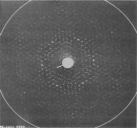
Highly ordered two-dimensional (2-D) crystals of Escherichia coli-expressed bacteriorhodopsin analog (e-bR) and its D96N variant (e-D96N) reconstituted in Halobacterium halobium lipids have been obtained by starting with the opsin protein purified in the denaturing detergent sodium dodecyl sulfate. These crystals embedded in glucose show electron diffraction in projection to better than 3.0 A at room temperature. This is the first instance that expressed bR or a variant has been crystallized in 2-D arrays showing such high order. The crystal lattice is homologous to that in wild-type bR (w-bR) in purple membranes (PM) and permit high resolution analyses of the structure of the functionally impaired D96N variant. The e-bR crystal is isomorphous to that in PM with an overall averaged fractional change of 12.7% (26-3.6-A resolution) in the projection structure factors. The projection difference Fourier map e-bR-PM at 3.6-A resolution indicates small conformational changes equivalent to movement of approximately < 7 C-atoms distributed within and in the neighborhood of the protein envelope. This result shows that relative to w-bR there are no global structural rearrangements in e-bR at this 3.6 A resolution level. The e-D96N crystal is isomorphous to the e-bR crystal with a smaller (9.2%) overall averaged fractional change in the structure factors. The significant structural differences between e-D96N and e-bR are concentrated at high resolution (5-3.6 A); however, these changes are small as quantified from the 3.6 A resolution e-D96N-e-bR Fourier difference map. The difference map showed no statistically significant peaks or valleys within 5 A in projection from the site of D96 substitution on helix C. Elsewhere within the protein envelope the integrated measure of peaks or valleys was < approximately 3 C-atom equivalents. Thus, our results show that for the isosteric substitution of Asp96 by Asn, the molecular conformation of bR in its ground state is essentially unaltered. Therefore, the known effect of D96N on the slowed M412 decay is not due to ground-state structural perturbations.
通过从在变性去污剂十二烷基硫酸钠中纯化的视蛋白开始,已经获得了在嗜盐菌脂质中重构的大肠杆菌表达的细菌视紫红质类似物(e-bR)及其D96N变体(e-D96N)的高度有序二维(2-D)晶体。这些包埋在葡萄糖中的晶体在室温下的投影中显示出优于3.0埃的电子衍射。这是首次将表达的细菌视紫红质或变体以显示如此高有序性的二维阵列形式结晶。晶格与紫色膜(PM)中野生型细菌视紫红质(w-bR)的晶格同源,并允许对功能受损的D96N变体的结构进行高分辨率分析。e-bR晶体与PM中的晶体同构,在投影结构因子中总体平均分数变化为12.7%(26 - 3.6埃分辨率)。3.6埃分辨率下的投影差分傅里叶图e-bR - PM表明,构象变化很小,相当于分布在蛋白质包膜内及其附近的约<7个碳原子的移动。该结果表明,相对于w-bR,在该3.6埃分辨率水平下e-bR中没有全局结构重排。e-D96N晶体与e-bR晶体同构,结构因子中的总体平均分数变化较小(9.2%)。e-D96N和e-bR之间的显著结构差异集中在高分辨率(5 - 3.6埃);然而,从3.6埃分辨率的e-D96N - e-bR傅里叶差分图量化来看,这些变化很小。差分图在从螺旋C上D96取代位点的投影中5埃范围内未显示出统计学上显著的峰或谷。在蛋白质包膜的其他地方,峰或谷的积分测量值<约3个碳原子当量。因此,我们的结果表明,对于天冬氨酸96被天冬酰胺的等排取代,细菌视紫红质在其基态的分子构象基本未改变。因此,已知的D96N对M412衰减减慢的影响不是由于基态结构扰动。