Glenn Biggs Institute for Neurodegenerative Disorders, University of Texas Health Science Center at San Antonio, San Antonio, TX, USA.
Research Imaging Institute, University of Texas Health Science Center at San Antonio, San Antonio, TX, USA.
J Alzheimers Dis. 2023;96(3):1267-1283. doi: 10.3233/JAD-230389.
Neuroimaging bears the promise of providing new biomarkers that could refine the diagnosis of dementia. Still, obtaining the pathology data required to validate the relationship between neuroimaging markers and neurological changes is challenging. Existing data repositories are focused on a single pathology, are too small, or do not precisely match neuroimaging and pathology findings.
The new data repository introduced in this work, the South Texas Alzheimer's Disease research center repository, was designed to address these limitations. Our repository covers a broad diversity of dementias, spans a wide age range, and was specifically designed to draw exact correspondences between neuroimaging and pathology data.
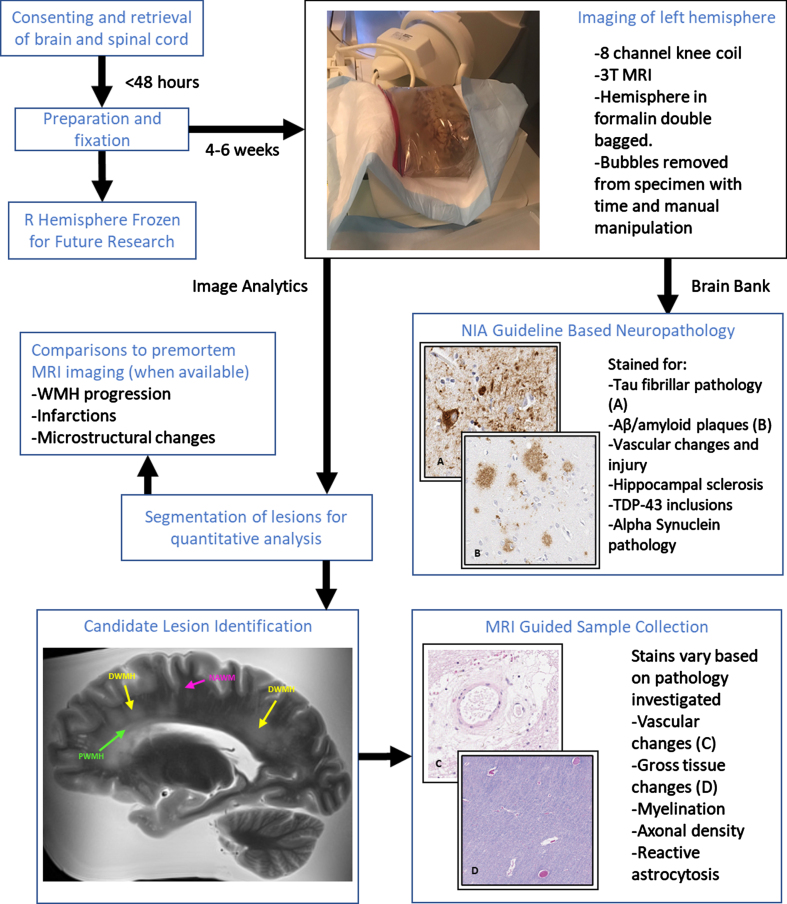
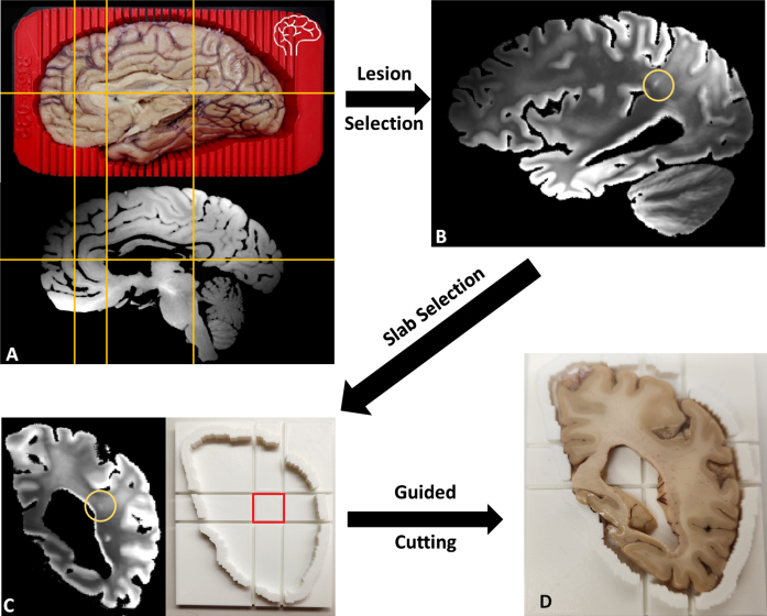
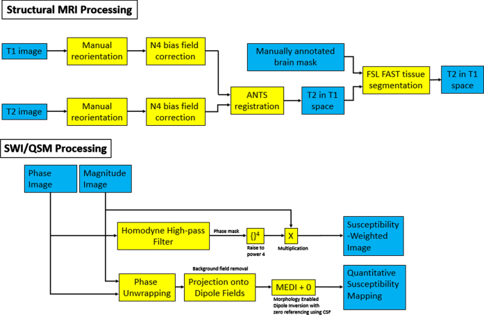
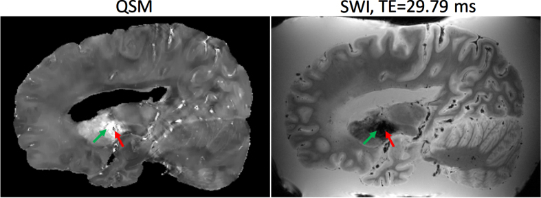
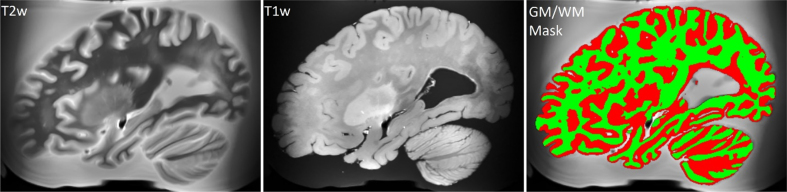
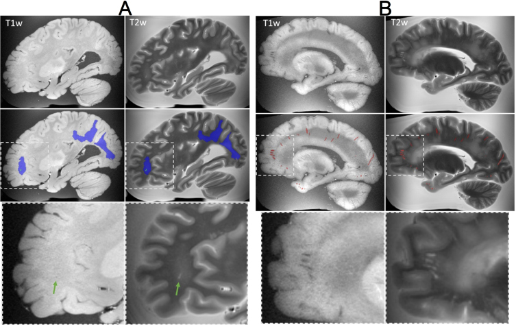
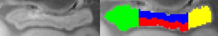
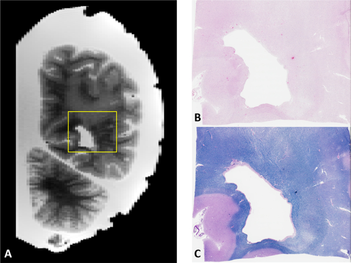
Using four different MRI sequences, we are reaching a sample size that allows for validating multimodal neuroimaging biomarkers and studying comorbid conditions. Our imaging protocol was designed to capture markers of cerebrovascular disease and related lesions. Quantification of these lesions is currently underway with MRI-guided histopathological examination.
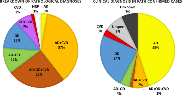
A total of 139 postmortem brains (70 females) with mean age of 77.9 years were collected, with 71 brains fully analyzed. Of these, only 3% showed evidence of AD-only pathology and 76% had high prevalence of multiple pathologies contributing to clinical diagnosis.
This repository has a significant (and increasing) sample size consisting of a wide range of neurodegenerative disorders and employs advanced imaging protocols and MRI-guided histopathological analysis to help disentangle the effects of comorbid disorders to refine diagnosis, prognosis and better understand neurodegenerative disorders.
神经影像学有望提供新的生物标志物,从而改善痴呆症的诊断。然而,获得验证神经影像学标志物与神经变化之间关系所需的病理学数据具有挑战性。现有的数据库专注于单一病理学,规模太小,或者与神经影像学和病理学发现不精确匹配。
本研究引入的新数据库——南德克萨斯州阿尔茨海默病研究中心数据库,旨在解决这些限制。我们的数据库涵盖了广泛的痴呆症,涵盖了广泛的年龄范围,并且专门设计用于在神经影像学和病理学数据之间建立精确对应关系。
我们使用四种不同的 MRI 序列,样本量达到了验证多模态神经影像学生物标志物和研究合并症的要求。我们的成像方案旨在捕获脑血管疾病和相关病变的标志物。目前正在通过 MRI 引导的组织病理学检查对这些病变进行定量分析。
共收集了 139 例尸检脑(70 名女性),平均年龄为 77.9 岁,其中 71 例脑得到了全面分析。其中,只有 3%的病例表现出 AD 型病理学证据,76%的病例存在多种导致临床诊断的病理学。
该数据库具有较大(且不断增加)的样本量,包含广泛的神经退行性疾病,并采用先进的成像方案和 MRI 引导的组织病理学分析,以帮助理清合并症的影响,从而改善诊断、预后,并更好地理解神经退行性疾病。