Department of Medical Biotechnology, School of Medicine, Zanjan University of Medical Sciences, Zanjan, Iran.
Nanotechnology Research Center, Ahvaz Jundishapur University of Medical Sciences, Ahvaz, Iran.
Oncol Res. 2023 Nov 15;32(1):101-125. doi: 10.32604/or.2023.044741. eCollection 2023.
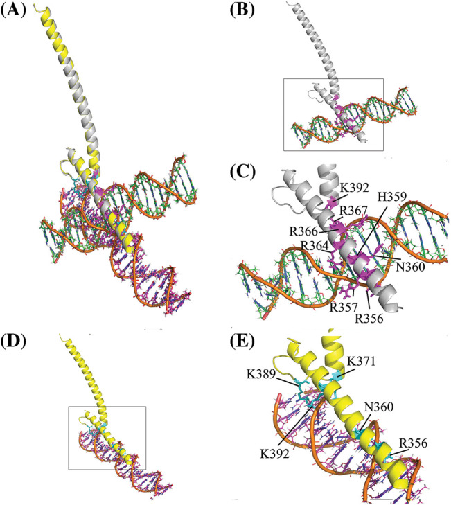
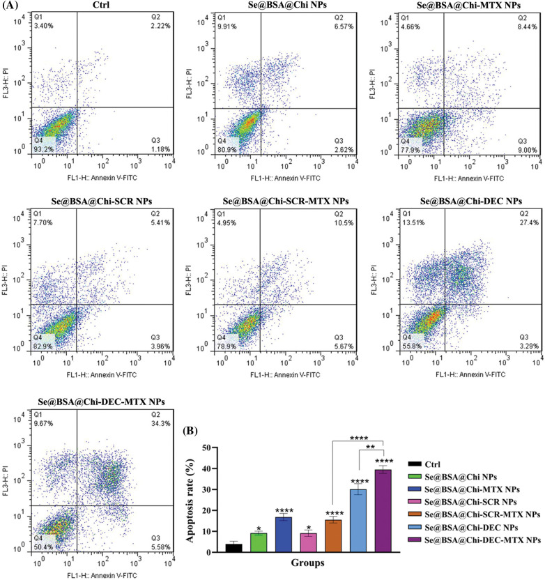
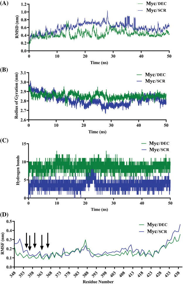
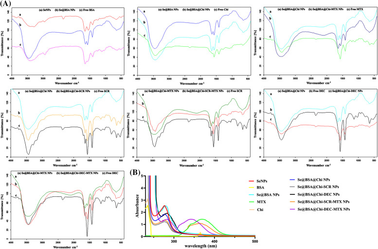
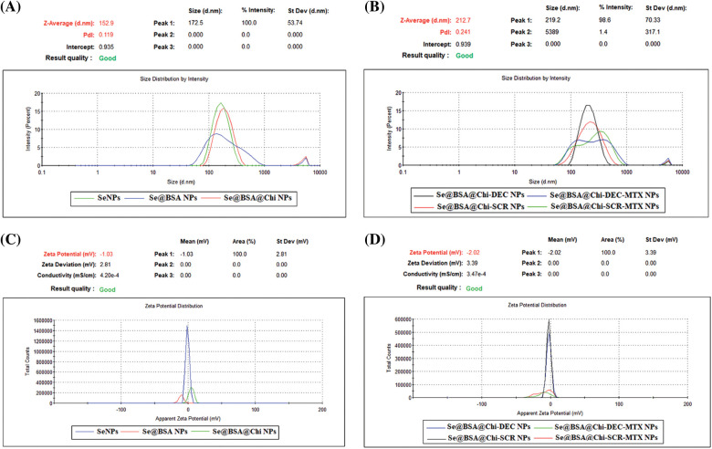
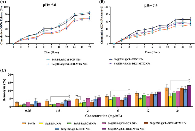
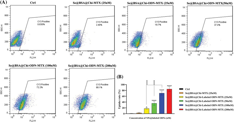
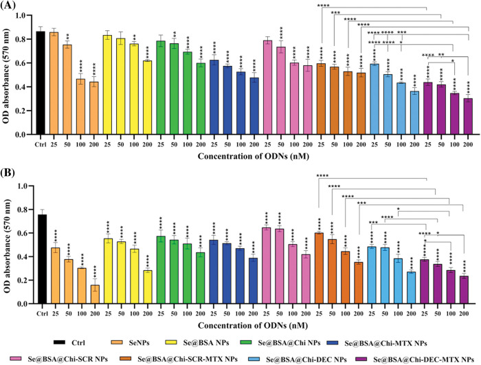
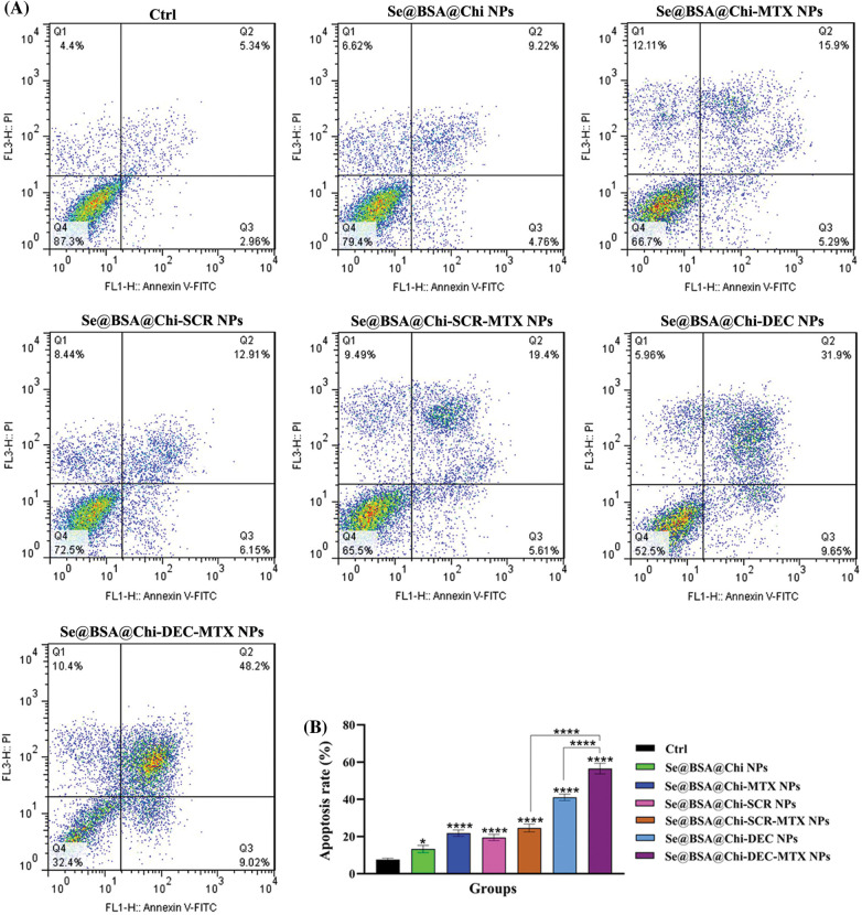
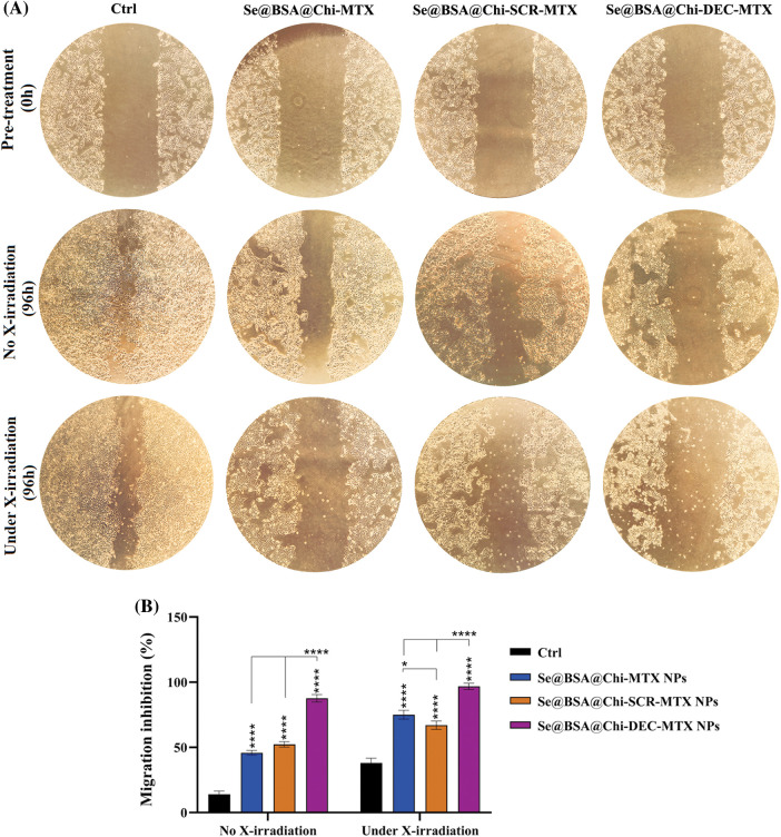
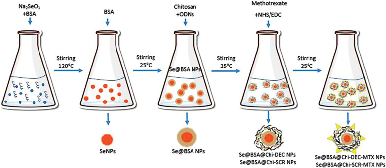
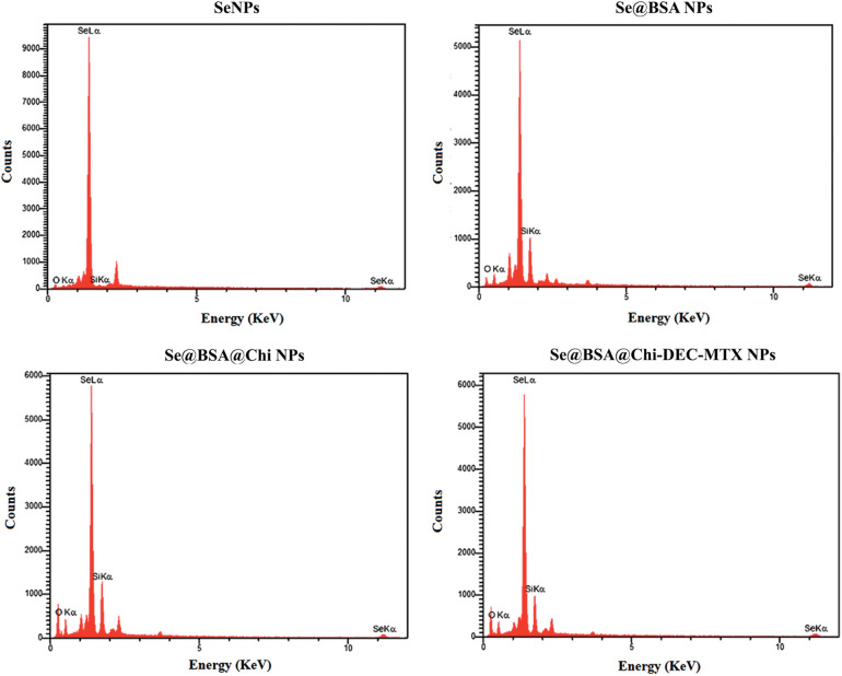
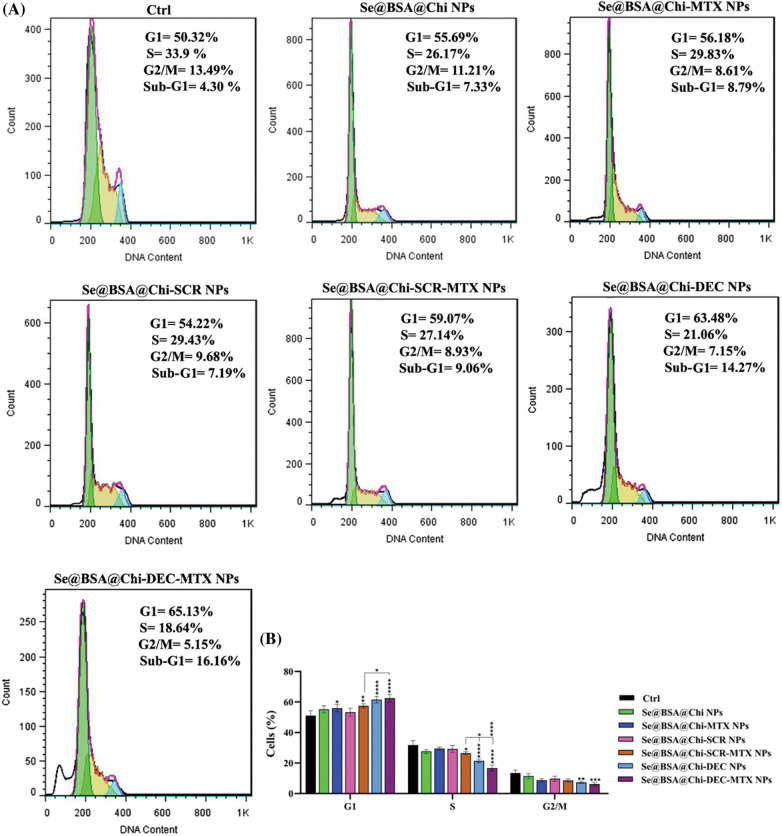
In the present study, we investigated the synergistic effects of targeted methotrexate-selenium nanostructure containing Myc decoy oligodeoxynucleotides along with X-irradiation exposure as a combination therapy on LNCaP prostate cancer cells. Myc decoy ODNs were designed based on the promoter of gene and analyzed by molecular docking and molecular dynamics assays. ODNs were loaded on the synthesized Se@BSA@Chi-MTX nanostructure. The physicochemical characteristics of nanostructures were determined by FTIR, DLS, UV-vis, TEM, EDX, release, and hemolysis tests. Subsequently, the cytotoxicity properties of them with and without X-irradiation were investigated by uptake, MTT, cell cycle, apoptosis, and scratch assays on the LNCaP cell line. The results of DLS and TEM showed negative charge (-9 mV) and nanometer size (40 nm) for Se@BSA@Chi-DEC-MTX NPs, respectively. The results of FTIR, UV-vis, and EDX showed the proper interaction of different parts and the correct synthesis of nanoparticles. The results of hemolysis showed the hemocompatibility of this nanoparticle in concentrations less than 6 mg/mL. The ODNs release from the nanostructures showed a pH-dependent manner, and the release rate was 15% higher in acidic pH. The targeted Se@BSA@Chi-labeled ODN-MTX NPs were efficiently taken up by LNCaP cells by targeting the prostate-specific membrane antigen (PSMA). The significant synergistic effects of nanostructure (containing MTX drug) treatment along with X-irradiation showed cell growth inhibition, apoptosis induction (~57%), cell cycle arrest (G2/M phase), and migration inhibition (up to 90%) compared to the control. The results suggested that the Se@BSA@Chi-DEC-MTX NPs can potentially suppress the cell growth of LNCaP cells. This nanostructure system can be a promising approach for targeted drug delivery and chemoradiotherapy in prostate cancer treatment.
在本研究中,我们研究了靶向甲氨蝶呤-硒纳米结构与 X 射线照射相结合作为联合治疗对 LNCaP 前列腺癌细胞的协同作用。Myc 诱饵寡脱氧核苷酸是根据基因启动子设计的,并通过分子对接和分子动力学分析进行了分析。将 ODN 加载到合成的 Se@BSA@Chi-MTX 纳米结构上。通过傅里叶变换红外光谱(FTIR)、动态光散射(DLS)、紫外-可见分光光度法(UV-vis)、透射电子显微镜(TEM)、能量色散 X 射线光谱(EDX)、释放和溶血试验来确定纳米结构的物理化学特性。随后,通过摄取、MTT、细胞周期、凋亡和划痕试验,研究了它们在有无 X 射线照射时对 LNCaP 细胞系的细胞毒性。DLS 和 TEM 的结果分别显示 Se@BSA@Chi-DEC-MTX NPs 的负电荷(-9 mV)和纳米尺寸(40 nm)。FTIR、UV-vis 和 EDX 的结果表明了不同部分的适当相互作用和纳米粒子的正确合成。溶血试验结果表明,该纳米颗粒在浓度低于 6 mg/mL 时具有血液相容性。ODNs 从纳米结构中的释放呈 pH 依赖性,在酸性 pH 下释放率提高 15%。靶向 Se@BSA@Chi 标记的 ODN-MTX NPs 通过靶向前列腺特异性膜抗原(PSMA)被 LNCaP 细胞有效摄取。与对照组相比,纳米结构(含 MTX 药物)治疗联合 X 射线照射表现出显著的协同作用,导致细胞生长抑制、凋亡诱导(约 57%)、细胞周期停滞(G2/M 期)和迁移抑制(高达 90%)。结果表明,Se@BSA@Chi-DEC-MTX NPs 可能抑制 LNCaP 细胞的生长。该纳米结构系统可能成为前列腺癌治疗中靶向药物递送和放化疗的有前途的方法。