Tian Jingxia, Wei Xiaoying, Zhang Weihua, Xu Aiguo
Department of Respiratory and Critical Care Medicine, First People's Hospital of Shangqiu, Shangqiu, China.
Department of Nephropathy of Rheumatology, First People's Hospital of Shangqiu, Shangqiu, China.
Front Bioeng Biotechnol. 2020 Nov 16;8:598997. doi: 10.3389/fbioe.2020.598997. eCollection 2020.
To investigate the effects of selenium nanoparticles (nano-Se) combined with radiotherapy on the proliferation, migration, invasion, and apoptosis of non-small cell lung cancer (NSCLC) A549 and NCI-H23 cells.
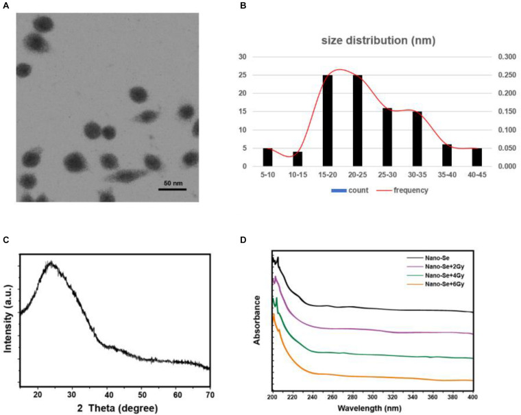
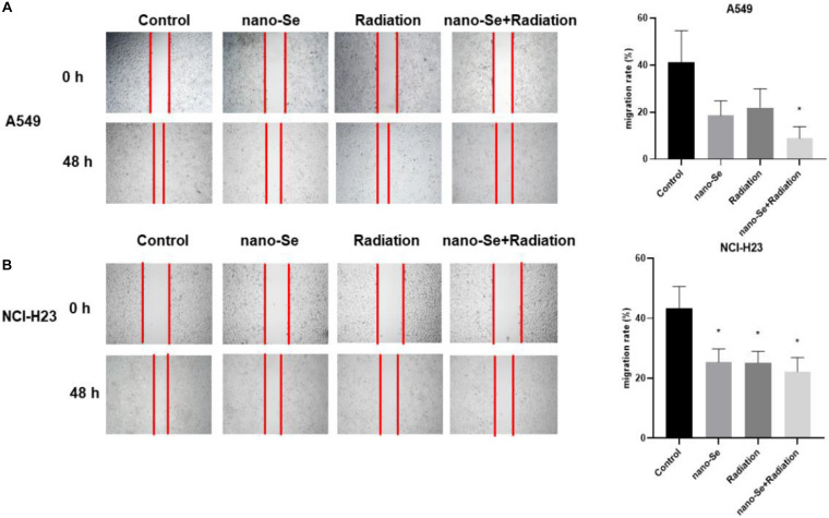
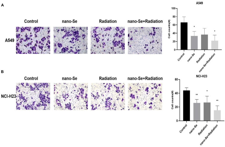
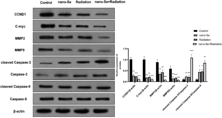
Nano-Se was synthesized and characterized by transmission electron microscope (TEM), X-ray diffractometer, and Ultraviolet-visible (UV)-Vis Spectroscopy, separately. The uptake of nano-Se by lung cancer cells was detected by flow cytometry. Cell counting kit-8 (CCK-8) method was used to detect the antiproliferative activity of nano-Se combined with radiotherapy. Wound healing tests and transwell assay were used to detect the migration and invasion ability of the cells. Annexin V-fluorescein isothiocyanate (FITC)/Propidium iodide (PI) staining by flow cytometry was used to detect apoptosis. The expression of Cyclin D1 (CCND1), c-Myc, matrix metalloproteinase 2 (MMP2), MMP9, cleaved Caspase-3, and cleaved Caspase-9 were detected by Western blot.
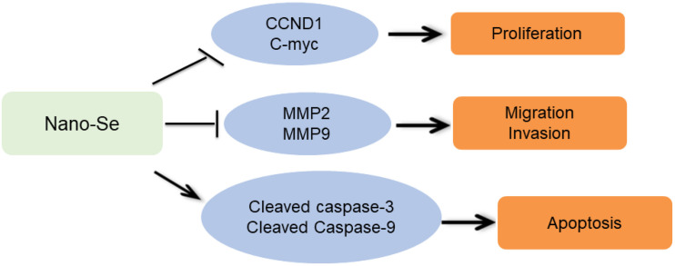
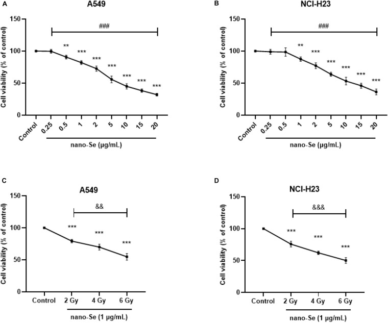
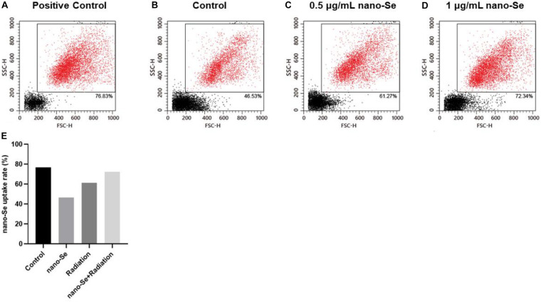
The average diameter of nano-Se was 24.39 nm and the wavelength of nano-Se increased with the increase of radiation dose under UV-Vis Spectroscopy. The uptake of nano-Se in lung cancer cells was increased with the increase of nano-Se concentration. The nano-Se combined with radiotherapy decreased the proliferation activity of NSCLC cell lines A549 and NCI-H23 in a dose-dependent manner (all < 0.05). Compared with the Control group, nano-Se combined with radiotherapy could significantly inhibit the migration and invasion of lung cancer cells (all < 0.05), and the effects of the combination of nano-Se and radiotherapy was better than that of a single application (all < 0.05). Furthermore, nano-Se combined with radiotherapy could induce apoptosis of lung cancer cells ( < 0.05) and nano-Se combined with radiotherapy could significantly inhibit the expression of proliferation-related proteins CCND1, c-Myc, invasion and migration-related proteins MMP2 and MMP9, but conversely promoted the expression of apoptosis-related proteins cleaved caspase-3 and cleaved caspase-9. Conclusion: This study found that nano-Se combined with radiotherapy plays an anti-cancer role in lung cancer cells by inhibiting cell proliferation, migration, and invasion, as well as inducing apoptosis, suggesting that nano-Se may be used as a radiosensitizer in the clinical treatment of lung cancer, but further research is still needed.
探讨纳米硒(nano-Se)联合放疗对非小细胞肺癌(NSCLC)A549和NCI-H23细胞增殖、迁移、侵袭及凋亡的影响。
分别采用透射电子显微镜(TEM)、X射线衍射仪和紫外可见(UV)-可见光谱对纳米硒进行合成与表征。通过流式细胞术检测肺癌细胞对纳米硒的摄取。采用细胞计数试剂盒-8(CCK-8)法检测纳米硒联合放疗的抗增殖活性。采用伤口愈合试验和Transwell实验检测细胞的迁移和侵袭能力。通过流式细胞术用膜联蛋白V-异硫氰酸荧光素(FITC)/碘化丙啶(PI)染色检测细胞凋亡。采用蛋白质免疫印迹法检测细胞周期蛋白D1(CCND1)、c-Myc、基质金属蛋白酶2(MMP2)、MMP9、裂解的半胱天冬酶-3和裂解的半胱天冬酶-9的表达。
纳米硒的平均直径为24.39 nm,在紫外可见光谱下,纳米硒的波长随辐射剂量的增加而增加。肺癌细胞对纳米硒的摄取随纳米硒浓度的增加而增加。纳米硒联合放疗以剂量依赖方式降低NSCLC细胞系A549和NCI-H23的增殖活性(均P<0.05)。与对照组相比,纳米硒联合放疗可显著抑制肺癌细胞的迁移和侵袭(均P<0.05),且纳米硒与放疗联合应用的效果优于单一应用(均P<0.05)。此外,纳米硒联合放疗可诱导肺癌细胞凋亡(P<0.05),纳米硒联合放疗可显著抑制增殖相关蛋白CCND1、c-Myc、侵袭和迁移相关蛋白MMP2和MMP9的表达,但相反地促进了凋亡相关蛋白裂解的半胱天冬酶-3和裂解的半胱天冬酶-9的表达。结论:本研究发现纳米硒联合放疗通过抑制细胞增殖、迁移和侵袭以及诱导凋亡在肺癌细胞中发挥抗癌作用,提示纳米硒可能作为肺癌临床治疗中的放射增敏剂,但仍需进一步研究。