Division of Nephrology, Renmin Hospital of Wuhan University, 238 Jiefang Rd, Wuhan, Hubei, 430060, China.
Cell Commun Signal. 2024 Jan 10;22(1):26. doi: 10.1186/s12964-023-01399-4.
Cardiolipin (CL) plays a critical role in maintaining mitochondrial membrane integrity and overall mitochondrial homeostasis. Recent studies have suggested that mitochondrial damage resulting from abnormal cardiolipin remodelling is associated with the pathogenesis of diabetic kidney disease (DKD). Acyl-coenzyme A:lyso-cardiolipin acyltransferase-1 (ALCAT1) was confirmed to be involved in the progression of Parkinson's disease, diet-induced obesity and other ageing-related diseases by regulating pathological cardiolipin remodelling. Thus, the purpose of this investigation was to determine the role of ALCAT1-mediated CL remodelling in DKD and to explore the potential underlying mechanism.
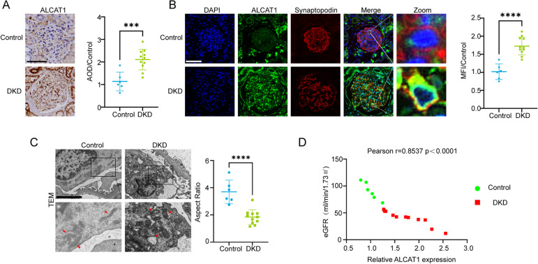
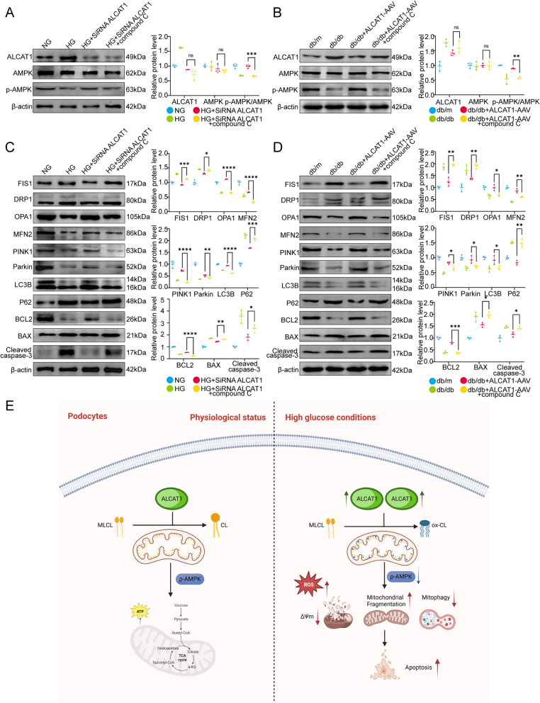
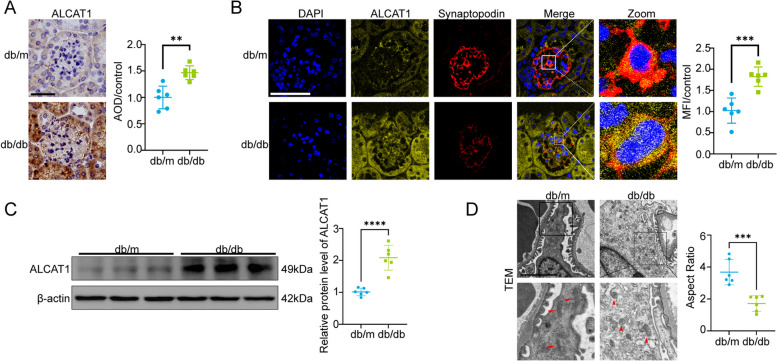
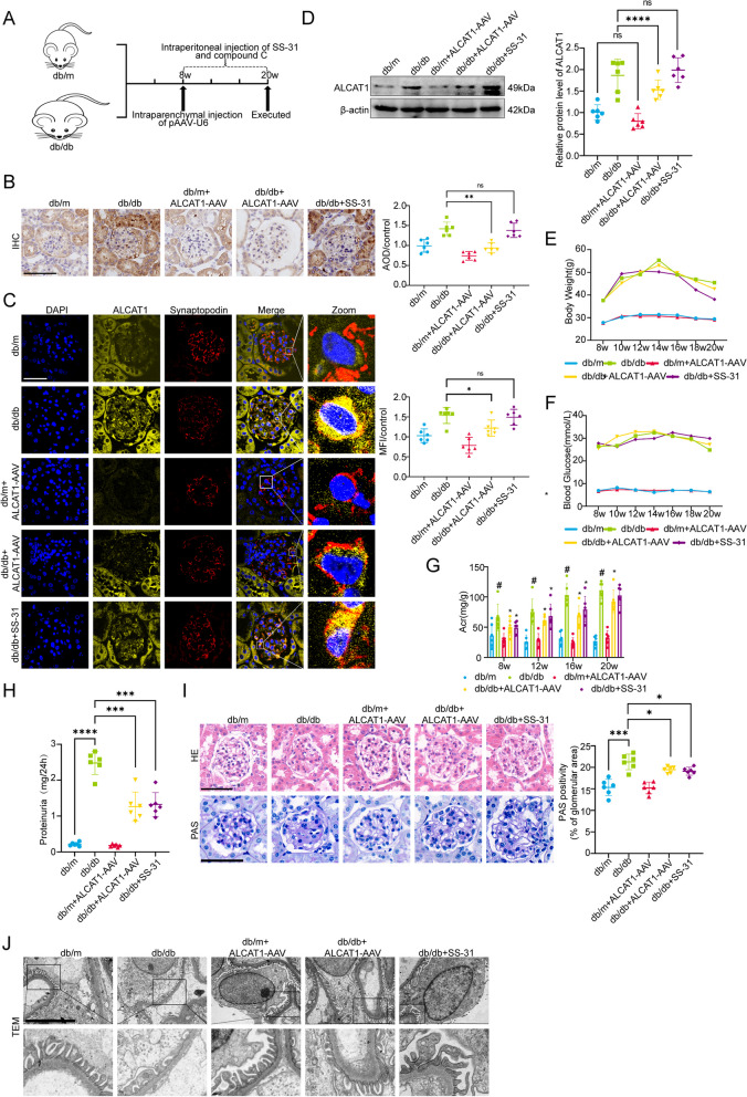
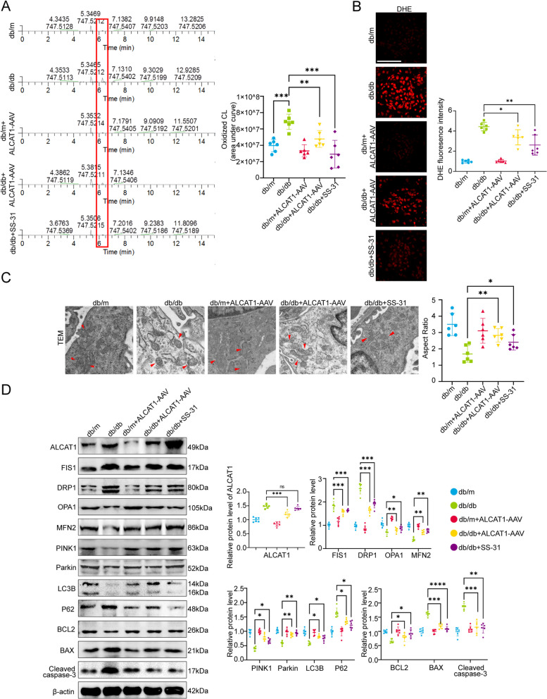
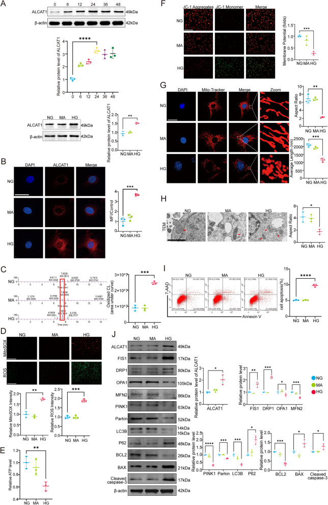
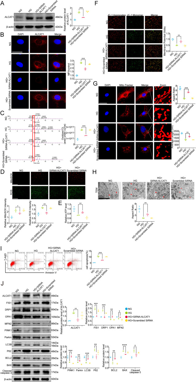
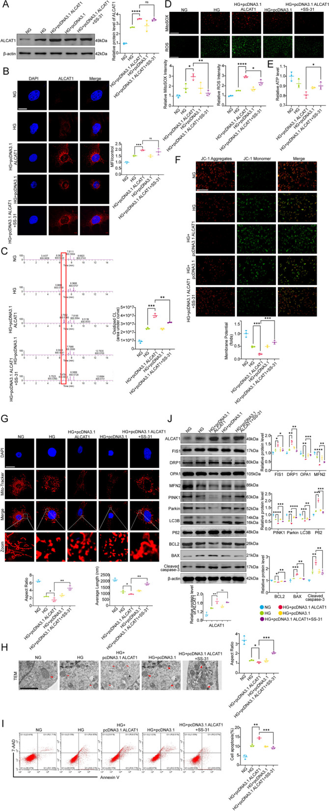
In vivo study, the mitochondrial structure was examined by transmission electron microscopy (TEM). The colocalization of ALCAT1 and synaptopodin was evaluated by double immunolabelling. Western blotting (WB) was performed to assess ALCAT1 expression in glomeruli. Lipidomics analysis was conducted to evaluate the composition of reconstructed cardiolipins. In vitro study, the lipidomics, TEM and WB analyses were similar to those in vivo. Mitochondrial function was evaluated by measuring the mitochondrial membrane potential (MMP) and the production of ATP and ROS.
Here, we showed that increased oxidized cardiolipin (ox-CL) and significant mitochondrial damage were accompanied by increased ALCAT1 expression in the glomeruli of patients with DKD. Similar results were found in db/db mouse kidneys and in cultured podocytes stimulated with high glucose (HG). ALCAT1 deficiency effectively prevented HG-induced ox-CL production and mitochondrial damage in podocytes. In contrast, ALCAT1 upregulation enhanced ox-CL levels and podocyte mitochondrial dysfunction. Moreover, treatment with the cardiolipin antioxidant SS-31 markedly inhibited mitochondrial dysfunction and cell injury, and SS-31 treatment partly reversed the damage mediated by ALCAT1 overexpression. We further found that ALCAT1 could mediate the key regulators of mitochondrial dynamics and mitophagy through the AMPK pathway.
Collectively, our studies demonstrated that ALCAT1-mediated cardiolipin remodelling played a crucial role in DKD, which might provide new insights for DKD treatment. Video Abstract.
心磷脂(CL)在维持线粒体膜完整性和整体线粒体动态平衡方面发挥着关键作用。最近的研究表明,异常的心磷脂重塑导致的线粒体损伤与糖尿病肾病(DKD)的发病机制有关。酰基辅酶 A:溶血心磷脂酰基转移酶-1(ALCAT1)通过调节病理性心磷脂重塑,被证实参与帕金森病、饮食诱导肥胖和其他与衰老相关疾病的进展。因此,本研究旨在探讨 ALCAT1 介导的心磷脂重塑在 DKD 中的作用及其潜在机制。
体内研究采用透射电子显微镜(TEM)观察线粒体结构。通过双重免疫标记评估 ALCAT1 和 synaptopodin 的共定位。Western blot(WB)检测肾小球中 ALCAT1 的表达。脂质组学分析评估重建心磷脂的组成。体外研究与体内研究相似,进行脂质组学、TEM 和 WB 分析。评估线粒体膜电位(MMP)和 ATP 和 ROS 的产生来评价线粒体功能。
我们发现,DKD 患者肾小球中氧化的心磷脂(ox-CL)增加和明显的线粒体损伤伴随着 ALCAT1 表达增加。db/db 小鼠肾脏和高糖(HG)刺激的培养足细胞中也发现了类似的结果。ALCAT1 缺乏可有效防止 HG 诱导的足细胞 ox-CL 产生和线粒体损伤。相反,ALCAT1 的上调增强了 ox-CL 水平和足细胞线粒体功能障碍。此外,用抗氧化剂 SS-31 处理可显著抑制线粒体功能障碍和细胞损伤,SS-31 处理部分逆转了由 ALCAT1 过表达介导的损伤。我们进一步发现,ALCAT1 可以通过 AMPK 通路调节线粒体动力学和线粒体自噬的关键调节剂。
综上所述,我们的研究表明,ALCAT1 介导的心磷脂重塑在 DKD 中起着关键作用,这可能为 DKD 的治疗提供新的思路。