Chair for High Performance Computing, Helmut-Schmidt-University Hamburg, Hamburg, Germany.
Center for Molecular Neurobiology (ZMNH), University Medical Center Hamburg-Eppendorf (UKE), Hamburg, Germany.
Brain Pathol. 2024 Sep;34(5):e13239. doi: 10.1111/bpa.13239. Epub 2024 Jan 11.
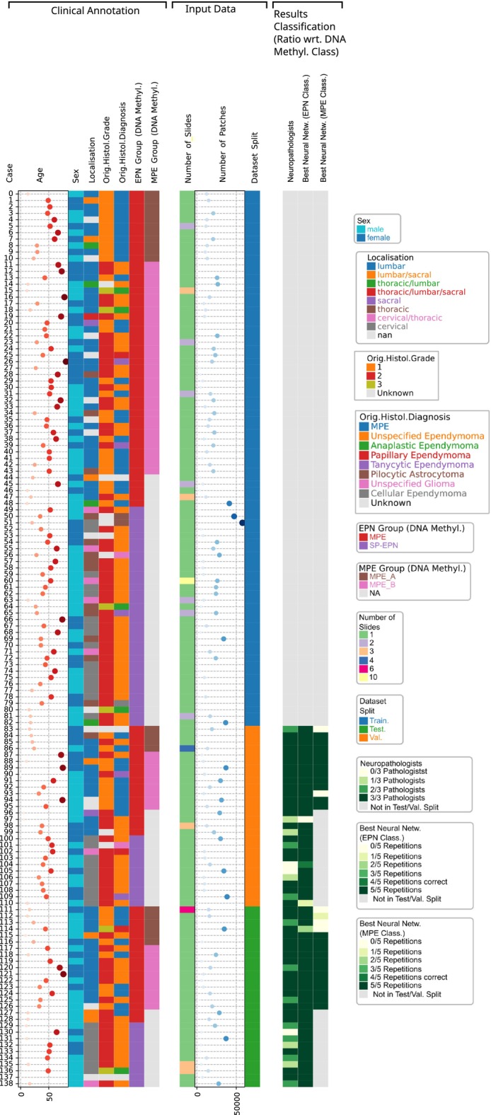
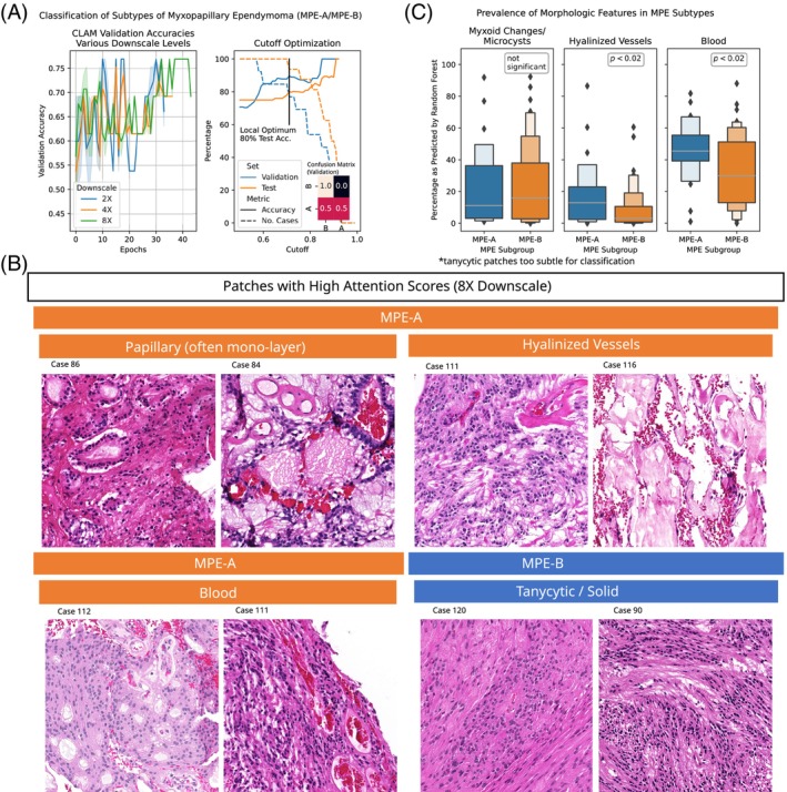
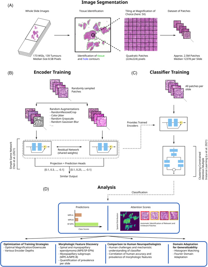
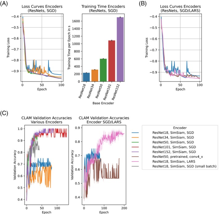
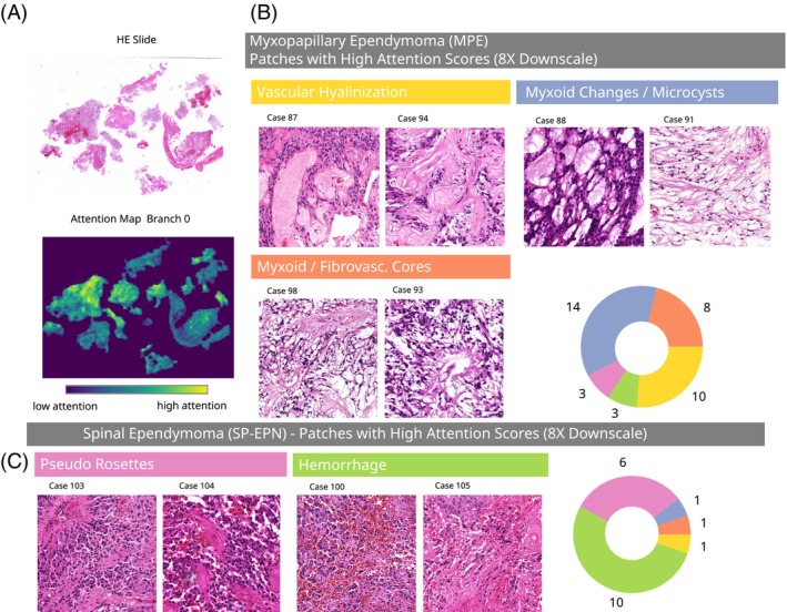
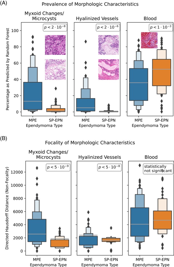
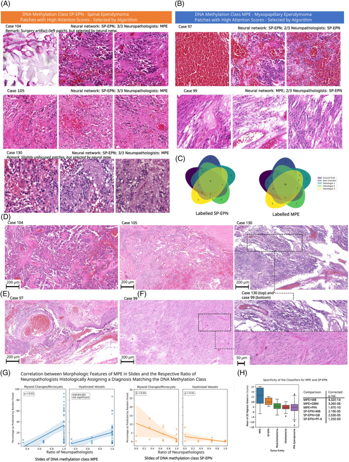
Based on DNA-methylation, ependymomas growing in the spinal cord comprise two major molecular types termed spinal (SP-EPN) and myxopapillary ependymomas (MPE(-A/B)), which differ with respect to their clinical features and prognosis. Due to the existing discrepancy between histomorphogical diagnoses and classification using methylation data, we asked whether deep neural networks can predict the DNA methylation class of spinal cord ependymomas from hematoxylin and eosin stained whole-slide images. Using explainable AI, we further aimed to prospectively improve the consistency of histology-based diagnoses with DNA methylation profiling by identifying and quantifying distinct morphological patterns of these molecular ependymoma types. We assembled a case series of 139 molecularly characterized spinal cord ependymomas (n = 84, n = 55). Self-supervised and weakly-supervised neural networks were used for classification. We employed attention analysis and supervised machine-learning methods for the discovery and quantification of morphological features and their correlation to the diagnoses of experienced neuropathologists. Our best performing model predicted the DNA methylation class with 98% test accuracy and used self-supervised learning to outperform pretrained encoder-networks (86% test accuracy). In contrast, the diagnoses of neuropathologists matched the DNA methylation class in only 83% of cases. Domain-adaptation techniques improved model generalization to an external validation cohort by up to 22%. Statistically significant morphological features were identified per molecular type and quantitatively correlated to human diagnoses. The approach was extended to recently defined subtypes of myxopapillary ependymomas (MPE-(A/B), 80% test accuracy). In summary, we demonstrated the accurate prediction of the DNA methylation class of spinal cord ependymomas (SP-EPN, MPE(-A/B)) using hematoxylin and eosin stained whole-slide images. Our approach may prospectively serve as a supplementary resource for integrated diagnostics and may even help to establish a standardized, high-quality level of histology-based diagnostics across institutions-in particular in low-income countries, where expensive DNA-methylation analyses may not be readily available.
基于 DNA 甲基化,脊髓内生长的室管膜瘤包含两种主要的分子类型,称为脊髓(SP-EPN)和黏液性乳头状室管膜瘤(MPE(-A/B)),它们在临床特征和预后方面存在差异。由于组织形态学诊断与基于甲基化数据的分类之间存在差异,我们想知道深度神经网络是否可以从苏木精和伊红染色的全切片图像预测脊髓室管膜瘤的 DNA 甲基化类型。我们使用可解释人工智能,进一步通过识别和量化这些分子室管膜瘤类型的不同形态模式,旨在前瞻性地提高基于组织学的诊断与 DNA 甲基化分析的一致性。我们收集了 139 例分子特征明确的脊髓室管膜瘤病例(n=84,n=55)。使用自我监督和弱监督神经网络进行分类。我们采用注意力分析和监督机器学习方法来发现和量化形态特征及其与经验丰富的神经病理学家诊断的相关性。我们表现最好的模型以 98%的测试准确性预测 DNA 甲基化类型,并使用自我监督学习来超越预训练的编码器网络(86%的测试准确性)。相比之下,病理学家的诊断与 DNA 甲基化类型仅在 83%的病例中相匹配。域自适应技术通过高达 22%的提高模型泛化到外部验证队列。每种分子类型都确定了具有统计学意义的形态特征,并与人类诊断定量相关。该方法扩展到最近定义的黏液性乳头状室管膜瘤亚型(MPE-(A/B),测试准确率为 80%)。总之,我们使用苏木精和伊红染色的全切片图像成功地预测了脊髓室管膜瘤(SP-EPN,MPE(-A/B))的 DNA 甲基化类型。我们的方法可能会成为综合诊断的补充资源,甚至有助于在整个机构中建立标准化的、高质量的基于组织学的诊断水平,尤其是在那些难以获得昂贵的 DNA 甲基化分析的低收入国家。