Department of Biomedical Engineering, University of California Irvine, Irvine, CA, USA.
Department of Radiological Sciences, School of Medicine, University of California Irvine, 839 Health Sciences Rd., Irvine, CA, 92617, USA.
J Transl Med. 2024 Jan 19;22(1):76. doi: 10.1186/s12967-024-04873-w.
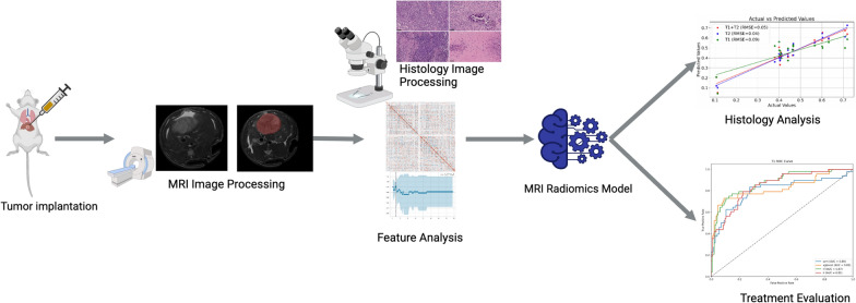
Hepatocellular carcinoma (HCC) is a common liver malignancy with limited treatment options. Previous studies expressed the potential synergy of sorafenib and NK cell immunotherapy as a promising approach against HCC. MRI is commonly used to assess response of HCC to therapy. However, traditional MRI-based metrics for treatment efficacy are inadequate for capturing complex changes in the tumor microenvironment, especially with immunotherapy. In this study, we investigated potent MRI radiomics analysis to non-invasively assess early responses to combined sorafenib and NK cell therapy in a HCC rat model, aiming to predict multiple treatment outcomes and optimize HCC treatment evaluations.
Sprague Dawley (SD) rats underwent tumor implantation with the N1-S1 cell line. Tumor progression and treatment efficacy were assessed using MRI following NK cell immunotherapy and sorafenib administration. Radiomics features were extracted, processed, and selected from both T1w and T2w MRI images. The quantitative models were developed to predict treatment outcomes and their performances were evaluated with area under the receiver operating characteristic (AUROC) curve. Additionally, multivariable linear regression models were constructed to determine the correlation between MRI radiomics and histology, aiming for a noninvasive evaluation of tumor biomarkers. These models were evaluated using root-mean-squared-error (RMSE) and the Spearman correlation coefficient.
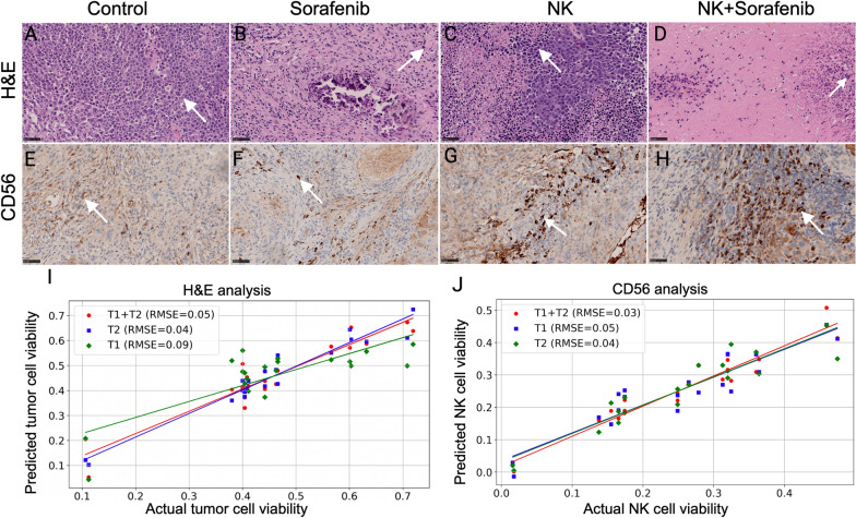
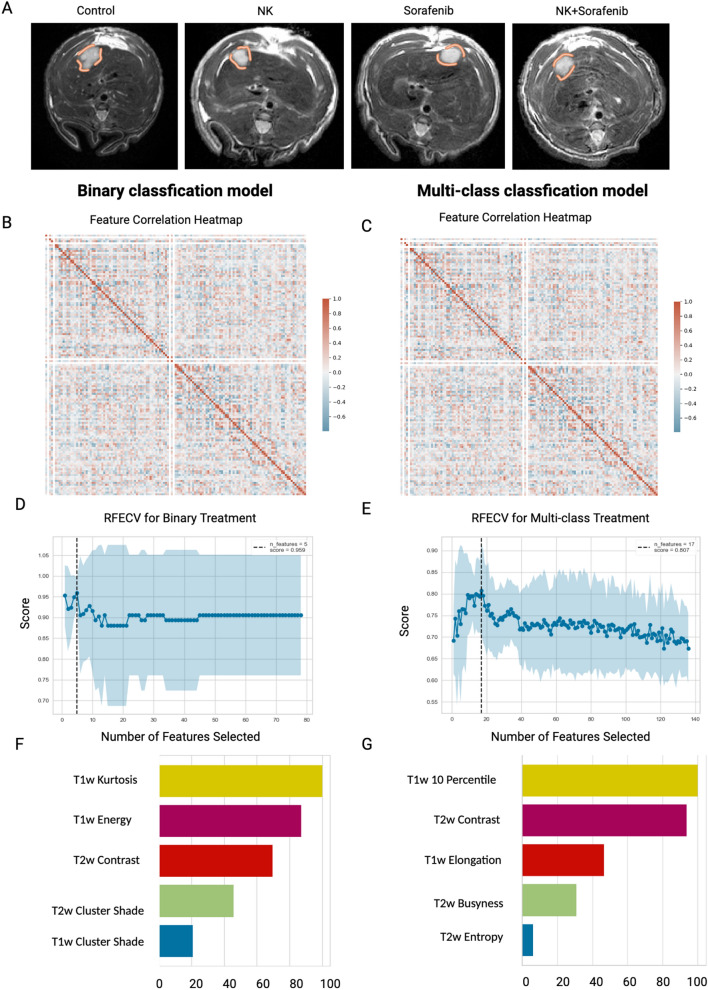
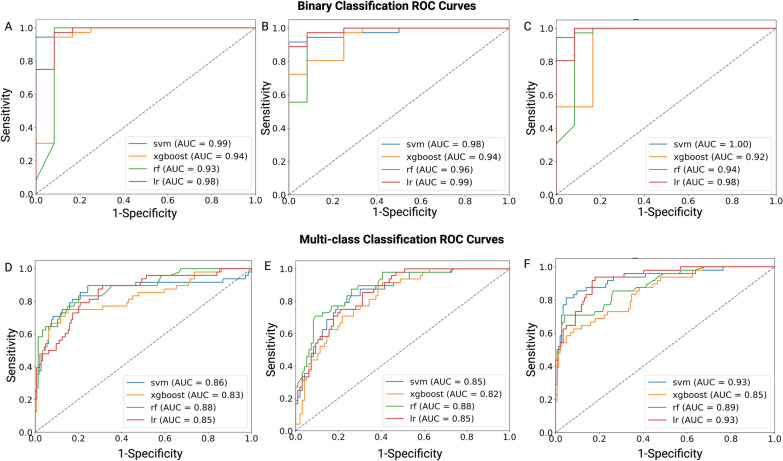
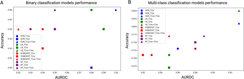
A total of 743 radiomics features were extracted from T1w and T2w MRI data separately. Subsequently, a feature selection process was conducted to identify a subset of five features for modeling. For therapeutic prediction, four classification models were developed. Support vector machine (SVM) model, utilizing combined T1w + T2w MRI data, achieved 96% accuracy and an AUROC of 1.00 in differentiating the control and treatment groups. For multi-class treatment outcome prediction, Linear regression model attained 85% accuracy and an AUC of 0.93. Histological analysis showed that combination therapy of NK cell and sorafenib had the lowest tumor cell viability and the highest NK cell activity. Correlation analyses between MRI features and histological biomarkers indicated robust relationships (r = 0.94).
Our study underscored the significant potential of texture-based MRI imaging features in the early assessment of multiple HCC treatment outcomes.
肝细胞癌(HCC)是一种常见的肝脏恶性肿瘤,治疗选择有限。先前的研究表明,索拉非尼和 NK 细胞免疫疗法的联合具有作为 HCC 治疗的有前途的方法的潜力。MRI 通常用于评估 HCC 对治疗的反应。然而,基于传统 MRI 的治疗效果指标不足以捕捉肿瘤微环境的复杂变化,尤其是免疫治疗。在这项研究中,我们研究了强大的 MRI 放射组学分析,以无创评估 HCC 大鼠模型中联合索拉非尼和 NK 细胞治疗的早期反应,旨在预测多种治疗结果并优化 HCC 治疗评估。
Sprague Dawley(SD)大鼠通过 N1-S1 细胞系进行肿瘤植入。在 NK 细胞免疫治疗和索拉非尼给药后,使用 MRI 评估肿瘤进展和治疗效果。从 T1w 和 T2w MRI 图像中提取、处理和选择放射组学特征。开发定量模型以预测治疗结果,并使用接收器操作特征(AUROC)曲线评估其性能。此外,构建多变量线性回归模型以确定 MRI 放射组学与组织学之间的相关性,旨在对肿瘤生物标志物进行无创评估。使用均方根误差(RMSE)和斯皮尔曼相关系数评估这些模型。
从 T1w 和 T2w MRI 数据中分别提取了 743 个放射组学特征。随后,进行了特征选择过程,以确定建模的五个特征子集。对于治疗预测,开发了四个分类模型。使用 T1w+T2w MRI 数据的支持向量机(SVM)模型实现了 96%的准确率和 1.00 的 AUROC,用于区分对照组和治疗组。对于多类治疗结果预测,线性回归模型达到了 85%的准确率和 0.93 的 AUC。组织学分析表明,NK 细胞和索拉非尼联合治疗具有最低的肿瘤细胞活力和最高的 NK 细胞活性。MRI 特征与组织学生物标志物之间的相关性分析表明存在很强的相关性(r=0.94)。
我们的研究强调了基于纹理的 MRI 成像特征在早期评估多种 HCC 治疗结果方面的重要潜力。